The Human Reproductive System: Anatomy, Hormones, and Development
An overview of the human reproductive system, detailing its anatomy, the roles of sex hormones, and the hormonal regulation through the HPG axis, including the menstrual cycle.
The human reproductive system is unique among all organ systems because it remains largely non-functional for the first 10 to 15 years of life. Unlike other systems that support the immediate survival of the individual, the reproductive system is designed for the continuation of the species, requiring the interaction of two complementary biological sexes to produce offspring. This process involves the production of specialized cells known as gametes, the secretion of sex hormones, and complex physiological cycles that prepare the body for potential pregnancy.
Genetic Sex and Early Development
The foundation of the reproductive system is laid at conception through the inheritance of sex chromosomes. While most females are genetically $46XX$ and most males are $46XY$, the biological determination of sex relies on a specific genetic trigger.
The presence of the SRY gene (Sex-determining Region of the Y chromosome) is the decisive factor in male development. If this gene is present, the individual develops as a male; in its absence, the individual develops a female phenotype, which is often considered the "default" genetic sex. It is important to distinguish between genetic sex and gender, as gender is a social construction that may not always align with an individual's biological sex.
Two major developmental milestones mark the lifespan of the reproductive system:
Puberty: The stage where reproductive organs reach sexual maturity and become capable of reproduction.
Menopause: The cessation of ovulation and menstruation, occurring when there has been no menstrual period for 12 consecutive months, due to the natural decline of hormone production in the ovaries.
The Male Reproductive System
The male reproductive system is designed for the production and delivery of male gametes, known as spermatozoa. The primary reproductive organs, or gonads, are the testes.
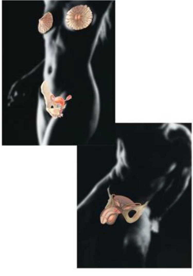
Diagrams of the female and male reproductive systems.
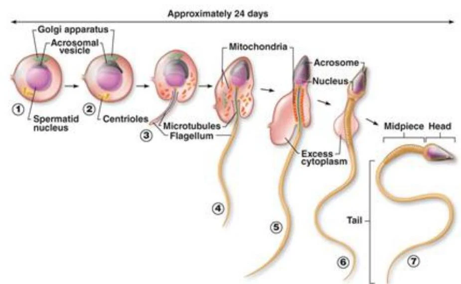
Within the testes, the process of spermatogenesis occurs inside the seminiferous tubules. Once produced, the spermatozoa move to the epididymides, where they undergo a maturation process necessary to acquire motility.
Diagram illustrating the process of spermatogenesis, showing the development of a sperm cell from a spermatid over approximately 24 days, highlighting key structures like the Golgi apparatus, mitochondria, nucleus, and flagellum.
The male system also includes several accessory structures:
Ducts: Pathways for the transport of secretions.
Glands: Including the prostate, bulbourethral, and seminal glands.
External Genitalia: The penis and scrotum.
An illustration of the male reproductive and urinary systems, labeling key anatomical structures such as the bladder, prostate, seminal vesicles, vas deferens, epididymis, and penis.
The Female Reproductive System
The female reproductive system is responsible for producing female gametes, called oocytes, and providing an environment for foetal development. The primary gonads are the ovaries.
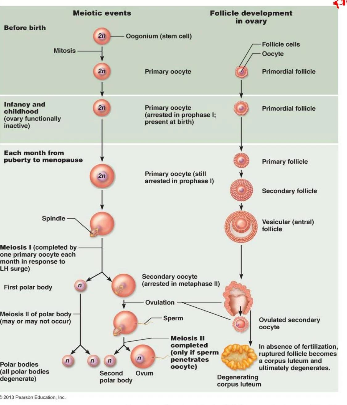
Unlike males, females are born with their complete complement of oocytes, which are housed within fluid-filled sacs called ovarian follicles. The ovarian cycle is the monthly series of events that leads to the maturation and ovulation (release) of an oocyte.
This diagram illustrates the process of oogenesis and follicle development in the ovary, detailing the stages from before birth through ovulation and potential fertilization or degeneration.
The female accessory organs include:
Uterine tubes: The site where fertilisation typically occurs.
Uterus: The site of foetal development.
Vagina: The passageway for menstruation and childbirth.
Hormonal Regulation: The HPG Axis
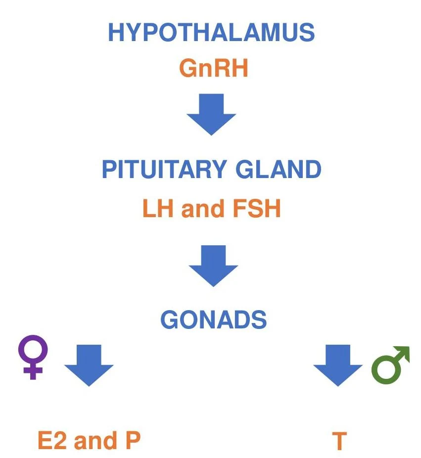
Reproduction is controlled by a complex feedback loop known as the Hypothalamic-Pituitary-Gonadal (HPG) axis. This system treats the hypothalamus, pituitary gland, and gonads as a single integrated entity.
This diagram illustrates the hormonal regulation of the reproductive system, starting with the hypothalamus releasing GnRH, which stimulates the pituitary gland to release LH and FSH. These hormones then act on the gonads, leading to the production of sex hormones like estrogen (E2), progesterone (P), and testosterone (T).
The process follows this hierarchy:
The hypothalamus secretes Gonadotropin-releasing hormone (GnRH).
GnRH triggers the anterior pituitary to release two key hormones: Luteinizing hormone (LH) and Follicle-stimulating hormone (FSH).
These hormones act on the gonads to stimulate the production of sex hormones.
Summary of Sex Hormones
Hormone
Primary Role in Males
Primary Role in Females
Testosterone (T)
Primary male hormone; promotes muscle mass, bone mass, and secondary sexual characteristics.
Produced in small amounts; aids in tissue maintenance.
Oestrogen (E2)
Exists in specific forms to support libido and erectile function.
Primary female hormone; promotes breast development and regulates the menstrual cycle.
Progesterone (P)
Influences spermiogenesis and counteracts oestrogen.
Maintains the uterine lining (endometrium) during pregnancy and supports lactation.
Exam Tip: Remember that LH has different "trigger" roles. In males, it stimulates Leydig cells to produce testosterone, whereas in females, an acute rise in LH is the specific trigger for ovulation.
The Menstrual Cycle
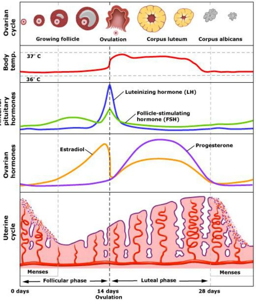
The menstrual cycle is a monthly process that governs the preparation and maintenance of the uterine lining. This cycle is driven by the fluctuating levels of ovarian hormones (oestrogen and progesterone).
The goal of this cycle is to create a thickened, nutrient-rich endometrium to support an embryo after implantation. If fertilisation does not occur, the hormone levels drop, causing the uterine lining to be shed, and the cycle begins again.
This diagram illustrates the hormonal and physiological changes during the human female reproductive cycle, including the ovarian cycle, anterior pituitary hormones, ovarian hormones, and uterine cycle.
Keep reading for free.
Create an account to finish this reading and make it part of your study routine.