The Human Reproductive System: Anatomy and Function
An overview of the human reproductive system, detailing the primary sex organs (gonads), accessory reproductive organs, and the processes of gamete production (spermatogenesis and oogenesis), fertilization, and development.
The human reproductive system is unique among the body's major systems because it is the only one that varies dramatically between biological sexes. While systems like the respiratory or circulatory systems are composed of organs found in every human, the reproductive system is specialized to facilitate the production of offspring through the creation of specialized cells and the secretion of sex hormones.
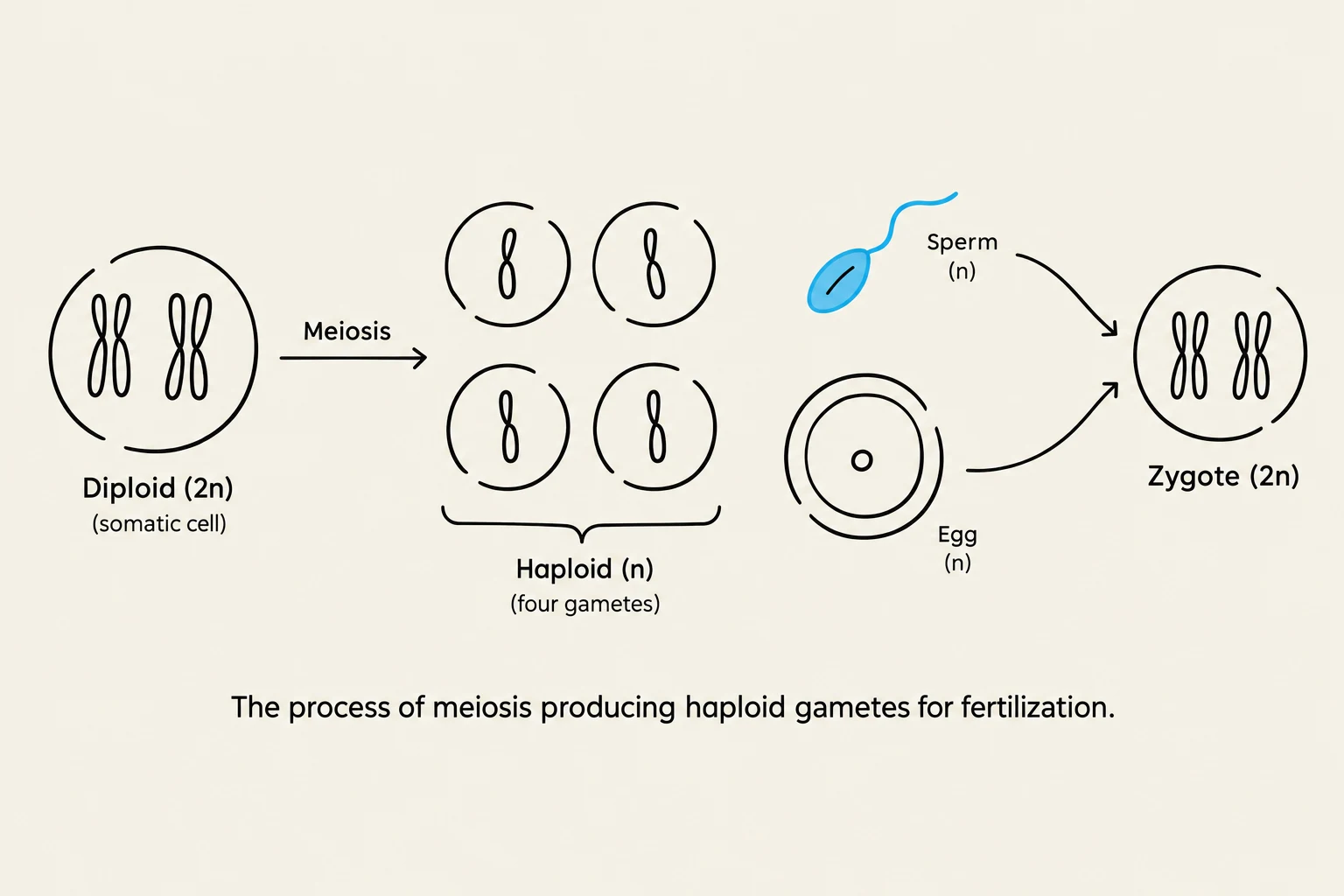
This diagram illustrates the biological process of meiosis, where a diploid somatic cell undergoes division to produce four haploid gametes. These gametes, a sperm and an egg, then fuse during fertilization to form a diploid zygote.
To understand how humans "make more humans," we must examine the system at two levels: the primary sex organs (gonads) and the accessory reproductive organs (ducts, glands, and genitalia).
The Foundation: Gonads and Gametes
At the heart of reproduction are the gonads, the primary sex organs responsible for two critical tasks: producing sex hormones and creating gametes.
Gametes are specialized reproductive cells produced through a process called meiosis. Unlike the standard somatic cells found throughout the rest of the body, which are diploid (containing two sets of chromosomes), gametes are haploid. This means they contain only half the genetic material of a normal body cell. This reduction is essential so that when two gametes unite, the resulting offspring has the correct diploid number of chromosomes.
The specific types of gametes produced depend on the sex of the individual:
Sperm cells: The male gametes.
Egg cells (Ova): The female gametes.
The union of a sperm cell and an egg cell is known as fertilization. This process results in the formation of a zygote, a single cell that marks the beginning of embryonic development and, eventually, a new human being.
Exam Tip: Mitosis vs. Meiosis
Remember that most cells in your body are produced via mitosis to create identical diploid cells. However, the reproductive system uses meiosis specifically to produce haploid gametes, ensuring genetic diversity and the correct chromosome count upon fertilization.
The Male Reproductive System
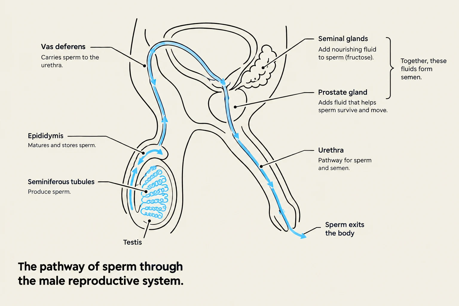
Diagram illustrating the pathway of sperm through the male reproductive system, including the vas deferens, epididymis, seminiferous tubules, testis, seminal glands, prostate gland, and urethra.
The male reproductive system is designed to produce, store, and deliver sperm cells into the female reproductive tract.
Primary Organs and Spermatogenesis
The male gonads are the testes. These are housed outside the main body cavity in a sac of skin called the scrotum. The scrotum serves a vital thermoregulatory function: the lower temperature found external to the body is ideal for sperm production.
Inside each testis, the tissue is divided into lobules containing coiled seminiferous tubules. It is within these tubules that spermatogenesis—the process of producing sperm—takes place.
The Duct System and Delivery
Once produced, sperm must travel through a series of ducts to reach the exit point:
Epididymis: Sperm move here from the testes to be stored until ejaculation.
Vas deferens & Ejaculatory duct: During ejaculation, sperm travel through these tubes.
Urethra: The final passage through which sperm exits the body.
The penis serves as the male copulatory organ, designed to deliver sperm into the female. It contains three cylindrical shafts of erectile tissue; during sexual arousal, these spaces engorge with blood, causing the organ to enlarge and harden (an erection) to enable penetration.
Accessory Glands and Semen
Sperm does not travel alone. It is mixed with fluids from several accessory glands to create semen. Semen is not the same as sperm; rather, it is the fluid medium that carries the sperm.
Seminal glands: Located on the surface of the bladder, they produce the majority of the semen volume. This fluid contains compounds that suppress the female immune response, destroy bacteria, and help sperm adhere to the vaginal walls to prevent drainage.
Prostate gland: This gland encircles the urethra and secretes fluid that activates the sperm.
Bulbo-urethral glands: These produce mucus to lubricate the glans penis during arousal.
The Female Reproductive System
Anatomy of the uterus and its three distinct layers.
The female reproductive system is anatomically more complex, involving the production of gametes, the potential for fetal development, and the production of milk.
Ovaries and Oogenesis
The female gonads are the ovaries. They are responsible for producing the egg cells (ova) and the essential sex hormones estrogen and progesterone.
The process of producing mature eggs is called oogenesis. Within the outer layer of the ovary (the cortex), specialized sacs called follicles house immature eggs known as oocytes. When a follicle matures, it undergoes ovulation, a process where the follicle bulges from the ovary surface and ejects the oocyte.
The Pathway to Fertilization and the Uterus
Following ovulation, the egg enters the uterine (fallopian) tubes. It is important to note that fertilization typically occurs within these tubes, not in the uterus itself.
If fertilization is successful, the resulting embryo moves into the uterus (the womb). The uterus is a highly specialized organ designed to receive, retain, and nourish a developing fetus. The uterine wall is composed of three distinct layers:
Perimetrium: The outer layer.
Myometrium: A thick, middle layer made of smooth muscle.
Endometrium: The inner lining of the uterine cavity.
External Genitalia and the Vagina
The vagina serves as the female copulatory organ, receiving the penis during intercourse. The external female genitalia are collectively referred to as the vulva, which includes the mons pubis, the labia majora and minora, the clitoris, and the vestibule.
The Mammary Glands
While often discussed separately, the mammary glands in the breasts are considered part of the female reproductive system due to their role in postnatal nourishment.
During pregnancy, these glands begin to produce milk. The anatomy of milk production is highly organized:
Alveoli: Small units within the lobules that actually produce the milk.
Lactiferous ducts: Tubes that transport the milk away from the alveoli.
Lactiferous sinus: A collection point where milk accumulates when a mother is nursing.
Through this intricate coordination of primary organs, accessory glands, and specialized ducts, the human body is capable of the complex biological feat of reproduction.
Keep reading for free.
Create an account to finish this reading and make it part of your study routine.