An overview of the human skeletal and muscular systems, their functions, classifications, and how they work together with the nervous system to enable movement. Covers joint types, movement terminology, muscle types, and the impact of exercise and immobility.
The human body relies on a complex interplay between the structural framework of the bones and the contractile power of the muscles to facilitate everything from delicate finger movements to powerful running strides. While the skeletal system provides the necessary scaffolding and protection, the muscular system acts as the engine that drives motion and maintains physiological stability. Understanding how these two systems integrate is fundamental to grasping human anatomy and kinesiology.
The Role of the Skeletal System
The skeletal system is far more than just a rigid frame; it is a dynamic organ system that performs several vital life-sustaining functions. Primarily, it supports the soft tissues of the body and protects crucial internal components, such as the brain and heart. Beyond protection, bones furnish the necessary surfaces for the attachment of muscles, tendons, and ligaments, which allows for coordinated movement.
On a microscopic and chemical level, the skeletal system serves as a reservoir. It provides storage areas for essential minerals and fat, and it is the primary site for the production of blood cells.
Classification of Bones by Shape
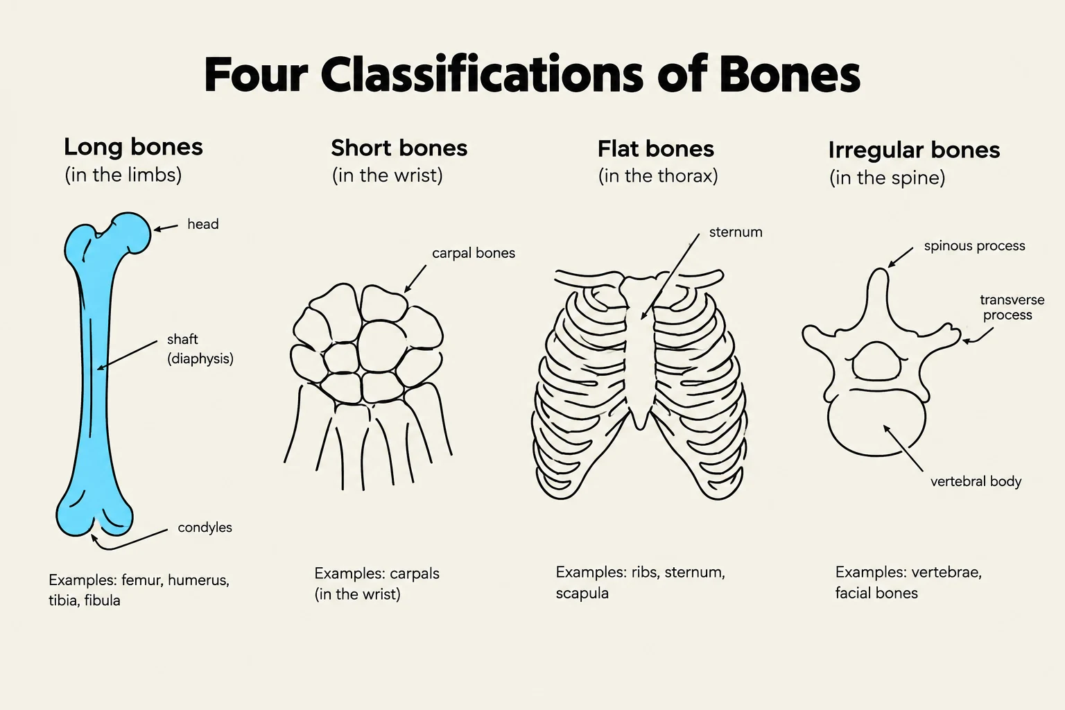
To better understand anatomy, bones are classified into four main categories based on their physical shape:
Long bones: Found in the upper and lower extremities, these bones contribute significantly to the body's height and length.
Short bones: Located in the wrist and ankle, these bones are essential for contributing to complex movements.
Flat bones: These include the ribs and skull bones; they are thin and serve to protect organs and contribute to the body's shape.
Irregular bones: This category includes all remaining bones that do not fit the other classifications, such as the jaw and the spinal column.
This illustration displays the four main classifications of bones: long bones, short bones, flat bones, and irregular bones, with examples and anatomical labels for each type.
Joints and Movement
Movement occurs at the junctions where bones meet, known as joints. While some joints are fixed, many are freely movable joints that allow for a wide range of motion. These include several specific types:
Ball and socket: Allows for multi-axial movement (e.g., hip or shoulder).
Hinge: Allows for movement in one plane (e.g., elbow).
Pivot: Allows for rotation around an axis.
Condyloid, Gliding, and Saddle joints: Various other configurations that facilitate specific directional movements.
Understanding Joint Movements
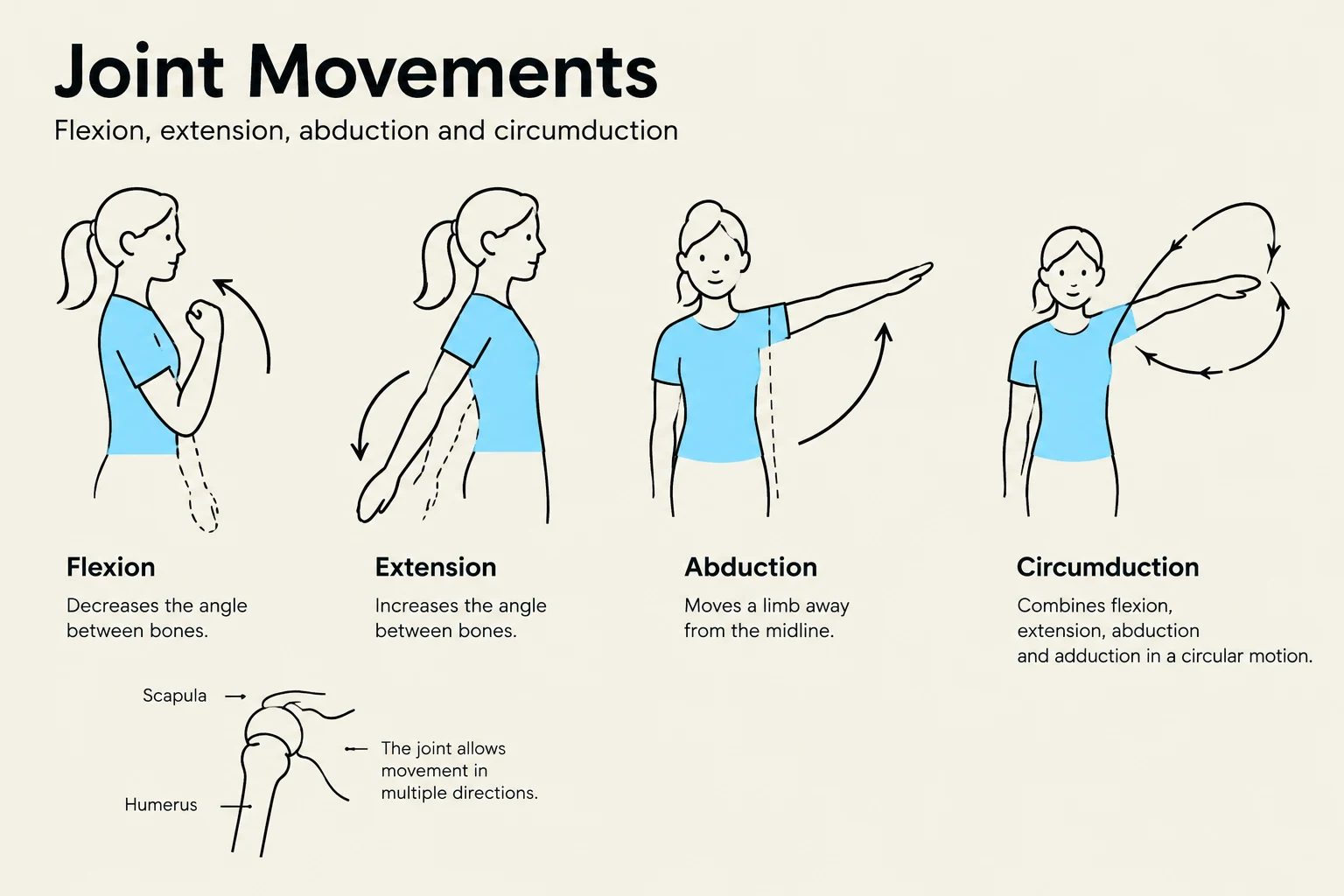
To describe how the body moves, clinicians use specific terminology to define the direction and nature of the motion:
Movement Type
Description
Flexion
The state of being bent; decreasing the angle between bones.
Extension
Increasing the angle between bones.
Abduction
Lateral movement of a body part away from the midline of the body.
Adduction
Movement of a body part toward the midline.
Circumduction
Moving the distal part of a limb in a circle while the proximal end remains fixed.
Dorsiflexion
The backward bending of the hand or foot.
Plantar flexion
The downward movement of the foot.
Exam Tip: If a question asks about moving a leg in a circular motion where the hip stays in place, the answer is circumduction, not abduction.
This illustration demonstrates four types of joint movements: flexion, extension, abduction, and circumduction, along with a diagram of the shoulder joint showing the humerus and scapula.
Muscle Functions and Types
If the bones are the levers, the muscles are the motors. There are three types of muscles in the human body:
Skeletal: Attached to bones; responsible for voluntary movement.
Cardiac: Found in the heart; responsible for pumping blood.
Smooth (or Visceral): Found in internal organs; responsible for involuntary functions.
The primary functions of muscles extend beyond simple motion. They are responsible for the maintenance of posture, providing support to the body, and heat production, which is vital for thermoregulation.
The Nervous System Connection
Muscles cannot move without instructions from the brain. This process involves a sophisticated communication loop through the nervous system:
Sensory Input: The afferent nervous system conveys information from peripheral receptors to the Central Nervous System (CNS).
Processing: The CNS processes this information.
Motor Output: The efferent neurons convey the response from the CNS to the skeletal muscles via the somatic nervous system, triggering a contraction.
This loop is supported by various reflexes, such as the proprioceptor (kinesthetic) sense, which helps the body understand its position in space, and stretch reflexes.
Exercise and Mobility
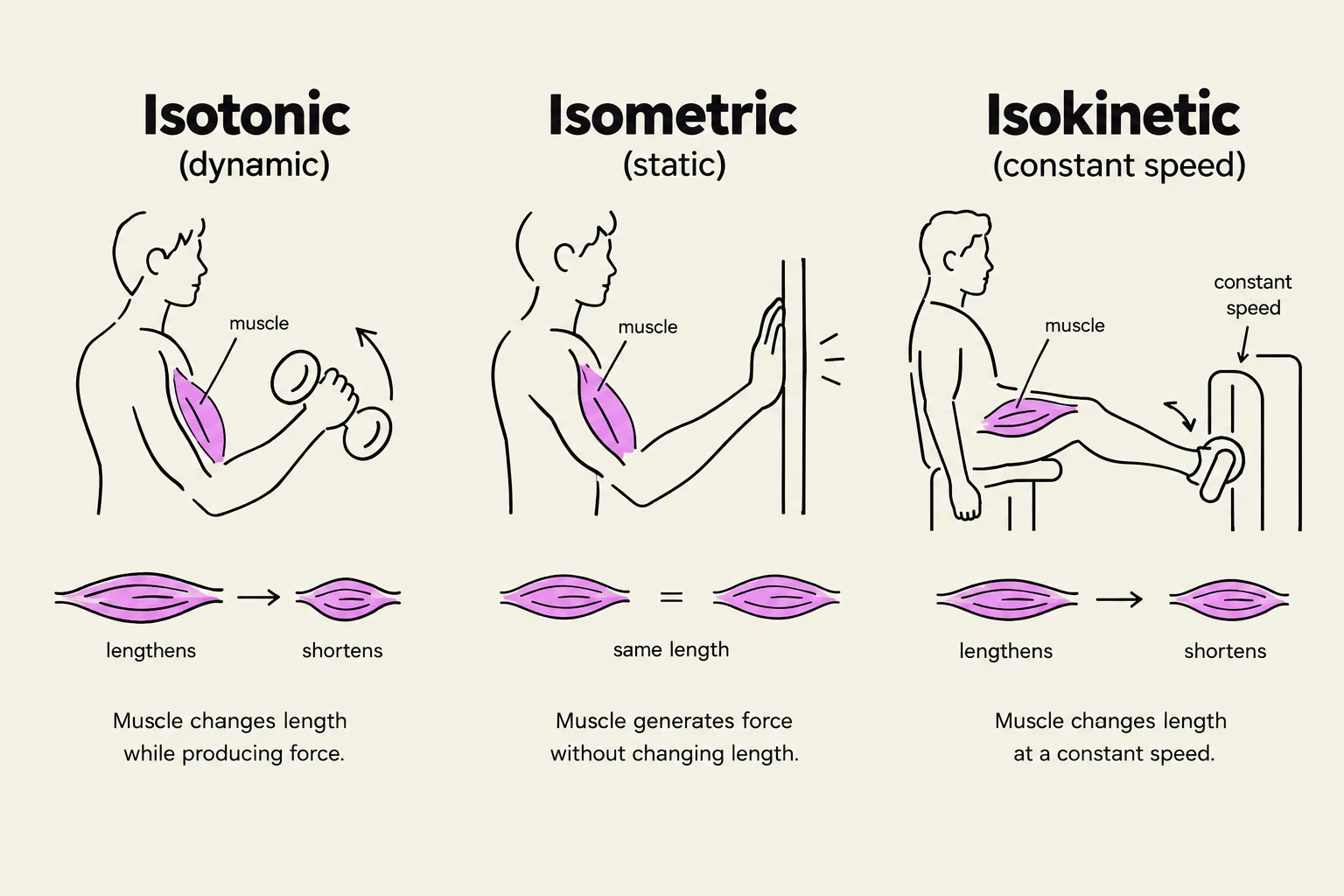
Maintaining the health of the musculoskeletal system requires regular physical activity. Exercise can be categorized into three types:
Isotonic: Involves muscle shortening and active movement (e.g., jogging).
Isometric: Involves muscle contraction without shortening the muscle length.
Isokinetic: Involves muscle contraction against a specific resistance.
Regular exercise provides systemic benefits, including increased muscle strength, improved bone density, and better cardiovascular efficiency. Conversely, immobility can lead to significant decline across the cardiovascular, respiratory, and metabolic systems.
Clinical Note: When assisting patients with mobility, healthcare workers must use proper body mechanics and assistive devices (like gait belts or mechanical lifts) to prevent injury, as uncoordinated lifts and repetitive tasks are major risk factors for back injuries.
This diagram illustrates three types of muscle contractions: isotonic (dynamic), isometric (static), and isokinetic (constant speed), explaining how muscle length changes or remains the same during force production.
Keep reading for free.
Create an account to finish this reading and make it part of your study routine.