An overview of the human motor system, including upper and lower motor neurons, descending pathways (pyramidal and extrapyramidal), and the role of the cerebellum in coordinating movement.
The human motor system is a sophisticated, hierarchical network designed to transform an abstract idea—such as "reach for that cup"—into a precise, coordinated physical movement. This process involves a seamless transition from high-level cognitive planning in the cortex to the execution of muscle contractions via the spinal cord. To understand how we move, we must examine the distinct roles of upper and lower motor neurons, the specific pathways they travel, and the regulatory influence of the cerebellum.
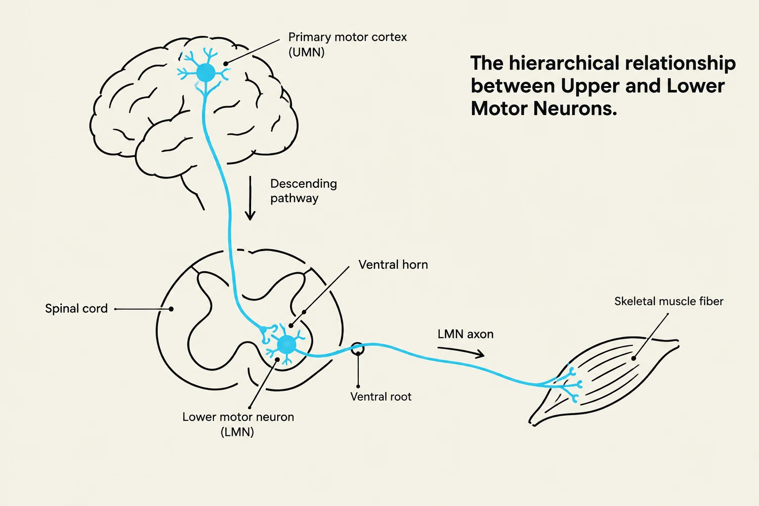
Diagram illustrating the hierarchical relationship between upper and lower motor neurons, showing the pathway from the primary motor cortex in the brain to a skeletal muscle fiber via the spinal cord.
The Hierarchical Organization: UMNs and LMNs
The motor system operates through a "top-down" hierarchy. At the highest levels, the cerebral cortex selects movement goals and strategies. These commands are then passed down through two primary types of neurons:
Upper Motor Neurons (UMNs)
UMNs are the "commanders" of the system. They originate in the primary motor cortex, the brainstem, or the vestibular nuclei. Their primary role is to initiate motor plans and send descending commands to the spinal cord or brainstem. Because they reside entirely within the Central Nervous System (CNS), they are responsible for regulating the excitability of the neurons below them.
Lower Motor Neurons (LMNs)
LMNs are known as the "Final Common Pathway". They originate in the spinal cord or brainstem and extend into the Peripheral Nervous System (PNS) to terminate directly on skeletal muscles. Because they are the final link in the chain, any signal intended to cause movement must pass through the LMN to reach the muscle. LMNs are highly myelinated to ensure rapid signal transmission to the target muscle.
Exam Tip: The "Final Common Pathway"
If a question asks which neuron is the "final common pathway" for all motor output to the muscles, the answer is always the Lower Motor Neuron (LMN).
Descending Pathways: Pyramidal vs. Extrapyramidal
Diagram illustrating the decussation (crossing over) of the lateral and anterior corticospinal tracts, which are major descending motor pathways from the brain to the spinal cord.
Motor pathways are categorized based on whether they are primarily involved in voluntary, skilled movements or involuntary, postural adjustments.
Pyramidal Pathways
These pathways are responsible for voluntary, purposeful, and complex movements. They include:
Corticospinal Tract: Controls the movements of the body and limbs.
Corticobulbar Tract: Controls the muscles of the head and neck by projecting to brainstem LMNs.
The Corticospinal Tract is further divided by where the axons decussate (cross the midline):
Lateral Corticospinal Tract: Decussates at the medulla. It governs the distal muscles of the extremities, allowing for "fractionated" movements—the ability to move individual muscles (like fingers) independently.
Anterior (Medial) Corticospinal Tract: Decussates at the level of the spinal cord. It governs the proximal muscles of the trunk and extremities via bilateral projection.
Extrapyramidal Pathways
These pathways manage non-voluntary, reflexive, or postural movements. They include:
Vestibulospinal Tracts: The medial tract orients head position, while the lateral tract helps maintain the center of gravity over the base of support.
Reticulospinal Tract: Involved in postural control and "antigravity" movements. Notably, it is the only medial tract that can influence distal extremity muscles, often producing abnormal muscle synergies in injury.
Rubrospinal Tract: Involved in postural control.
Somatotopic Organization in the Spinal Cord
Within the spinal cord, LMN cell bodies are not scattered randomly; they are clustered in specific groups called LMN pools located in Lamina IX of the ventral horn. These pools follow strict organizational rules:
Distal to Proximal Rule: Cell bodies for distal muscles (e.g., fingers) are located in the lateral ventral horn, whereas cell bodies for proximal/axial muscles (e.g., trunk/pelvis) are in the medial ventral horn.
Flexor-Extensor Rule: Cell bodies for flexor muscles are located posteriorly within the ventral horn, while extensor muscle cell bodies are located anteriorly.
Clinical Correlation: Myotomes
Understanding which spinal levels control which muscles is vital for clinical assessment:
C5: Shoulder abduction (deltoid)
C6: Elbow flexion (biceps)
C7: Elbow extension (triceps)
C8: Wrist flexion
T1: Finger abduction
L2: Hip flexion (iliopsoas)
L4: Knee extension (quadriceps)
L5: Dorsiflexion (tibialis anterior)
S1: Plantar flexion (gastrocnemius)
The Cerebellum: The Great Comparator
The cerebellum does not initiate movement, but it is essential for coordinating it. It acts as a "comparator," constantly evaluating the intended movement against the actual movement.
To do this, it processes two distinct information streams:
Intended Movement (Efference Copy): The cerebellum receives a "blueprint" of the motor plan via the Ventral (Anterior) Spinocerebellar Tract (VSCT) for the legs and the Rostral Cerebellar Tract (RSCT) for the arms.
Actual Movement (Proprioception): The cerebellum receives high-fidelity sensory feedback regarding what is actually happening via the Dorsal (Posterior) Spinocerebellar Tract (DSCT) for the legs and the Cuneocerebellar Tract (CCT) for the arms.
Because these pathways are typically unilateral (they do not cross or they cross twice), cerebellar lesions result in ipsilateral signs (deficits on the same side as the lesion).
Clinical Presentation: UMN vs. LMN Lesions
Distinguishing between an Upper Motor Neuron lesion and a Lower Motor Neuron lesion is a fundamental clinical skill.
Clinical Note: Spasticity vs. Hypotonia
In an acute UMN injury, you may initially see "spinal shock," characterized by temporary hypotonia and hyporeflexia. However, as the injury progresses to the later stage, the loss of descending regulation leads to the classic "positive" signs: hyperreflexia and spasticity (velocity-dependent hypertonia).
Keep reading for free.
Create an account to finish this reading and make it part of your study routine.