Explore the human digestive system, detailing the processes of mechanical and chemical digestion, the structure and function of the alimentary canal and accessory organs, and the roles of various secretions and enzymes in breaking down food for absorption.
The digestive system is a complex network of organs designed to perform a singular, vital task: converting the food we eat into forms that cell membranes can actually absorb. This process, known as digestion, involves a coordinated effort between a continuous muscular tube and several specialized organs that provide necessary chemical secretions.
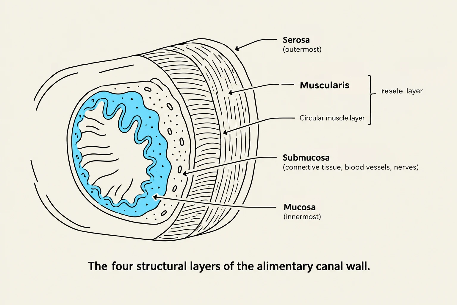
The four structural layers of the alimentary canal wall: Serosa (outermost), Muscularis (including the circular muscle layer), Submucosa (connective tissue, blood vessels, nerves), and Mucosa (innermost).
Understanding Digestion: Mechanical vs. Chemical
Digestion is not a single event but a combination of two distinct processes:
Mechanical Digestion: This involves the physical breakdown of large food pieces into smaller ones. While this increases the surface area for enzymes to work, it does not change the actual chemical composition of the food.
Chemical Digestion: This process uses enzymes to break down food particles into simpler chemical substances.
To achieve these goals, the body utilizes two main components: the alimentary canal (the food passageway) and the accessory organs (which secrete substances into the canal but do not have food passing through them).
The Alimentary Canal: Structure and Movement
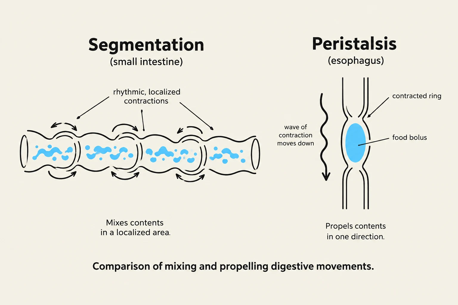
Comparison of mixing and propelling digestive movements: Segmentation in the small intestine mixes contents in a localized area through rhythmic, localized contractions, while peristalsis in the esophagus propels contents in one direction via a wave of contraction.
The alimentary canal is a muscular tube approximately 8 meters long, extending from the mouth to the anus. It includes the mouth, pharynx, esophagus, stomach, small intestine, large intestine, and anal canal.
The Four Layers of the Canal Wall
Regardless of the specific organ, the wall of the alimentary canal is generally composed of four distinct layers:
The muscularis layer is responsible for two primary types of movement:
Mixing Movements: Rhythmic contractions of small muscle sections that do not move material in one direction, such as segmentation in the small intestine or churning in the stomach.
Propelling Movements: One-way movement, most notably peristalsis, where a ring of contraction progresses down the tube in a wave-like motion.
Exam Tip: Remember that the autonomic nervous system regulates these movements. Parasympathetic impulses increase digestive activity (secretion and motility), while sympathetic impulses inhibit them.
The Beginning of the Journey: Mouth and Salivary Glands
The mouth is the first part of the alimentary canal, where mastication (chewing) begins the mechanical breakdown of food. The mouth is surrounded by the lips, cheeks, tongue, and palate.
Teeth: These are the hardest structures in the body and are used for biting (incisors), grasping/tearing (canines), and grinding (molars and premolars).
Tongue: A muscular organ that mixes food with saliva and contains taste buds (papillae).
Salivary Glands: Three major pairs—the parotid, submandibular, and sublingual glands—secrete saliva.
Saliva is crucial because it moistens food, contains bicarbonate ions to protect teeth from acid, and provides salivary amylase, an enzyme that begins the chemical digestion of carbohydrates by splitting starch into disaccharides.
Transport: Pharynx and Esophagus
While the pharynx and esophagus do not participate in actual digestion, they are essential for swallowing.
The Pharynx: A cavity posterior to the mouth that acts as a passageway for both air and food.
The Esophagus: A 25 cm muscular tube that uses peristalsis to move a food bolus to the stomach.
The swallowing reflex is a highly coordinated event. To prevent food from entering the respiratory tract, the soft palate and uvula rise to protect the nasal cavity, and the epiglottis closes off the top of the larynx to protect the trachea.
The Stomach: Chemical Processing and Mixing
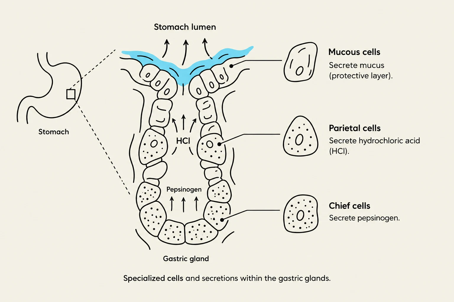
Diagram illustrating specialized cells within gastric glands and their secretions, including mucous cells, parietal cells secreting hydrochloric acid (HCl), and chief cells secreting pepsinogen.
The stomach is a J-shaped organ that receives food from the esophagus and mixes it with gastric juice to create a semifluid paste called chyme.
Gastric Secretions
The stomach's gastric glands contain specialized cells that produce "gastric juice":
Chief Cells: Secrete pepsinogen, an inactive enzyme that is converted into pepsin by hydrochloric acid to digest proteins.
Parietal Cells: Produce hydrochloric acid (HCl), which provides the acidic environment needed for pepsin, and intrinsic factor, which is required for Vitamin B12 absorption.
Mucous Cells: Secrete mucus to protect the stomach lining from the highly acidic environment.
Clinical Note:Heartburn (esophagitis) occurs when stomach contents reflux into the esophagus, causing inflammation. This is often caused by eating too quickly or consuming caffeine, nicotine, or alcohol.
Accessory Organs: Pancreas, Liver, and Gallbladder
As chyme enters the small intestine (duodenum), accessory organs release secretions to complete the digestive process.
The Pancreas
The pancreas acts as both an endocrine and exocrine gland. Its exocrine function involves secreting pancreatic juice into the duodenum via the pancreatic duct. This juice contains:
Pancreatic amylase (carbohydrates)
Pancreatic lipase (fats)
Trypsin, chymotrypsin, and carboxypeptidase (proteins)
Bicarbonate ions to neutralize acidic chyme.
The Liver and Gallbladder
The liver is the largest internal organ and performs hundreds of functions, including detoxification, protein metabolism, and storage of vitamins. Its primary role in digestion is the production of bile.
Bile is often stored and concentrated in the gallbladder. When fat enters the duodenum, the hormone cholecystokinin (CCK) is released, stimulating the gallbladder to contract and release bile into the digestive tract.
Keep reading for free.
Create an account to finish this reading and make it part of your study routine.