


The human eye is one of the most complex sensory organs in the body, acting as a biological camera that captures light and converts it into electrical signals for the brain to interpret. To understand how vision works, one must first master the gross anatomy of the eyeball. The eye is not merely a single unit but a sophisticated arrangement of specialized layers, chambers, and fluids designed to refract, focus, and protect light as it travels toward the retina.
The Three Tunics of the Eye
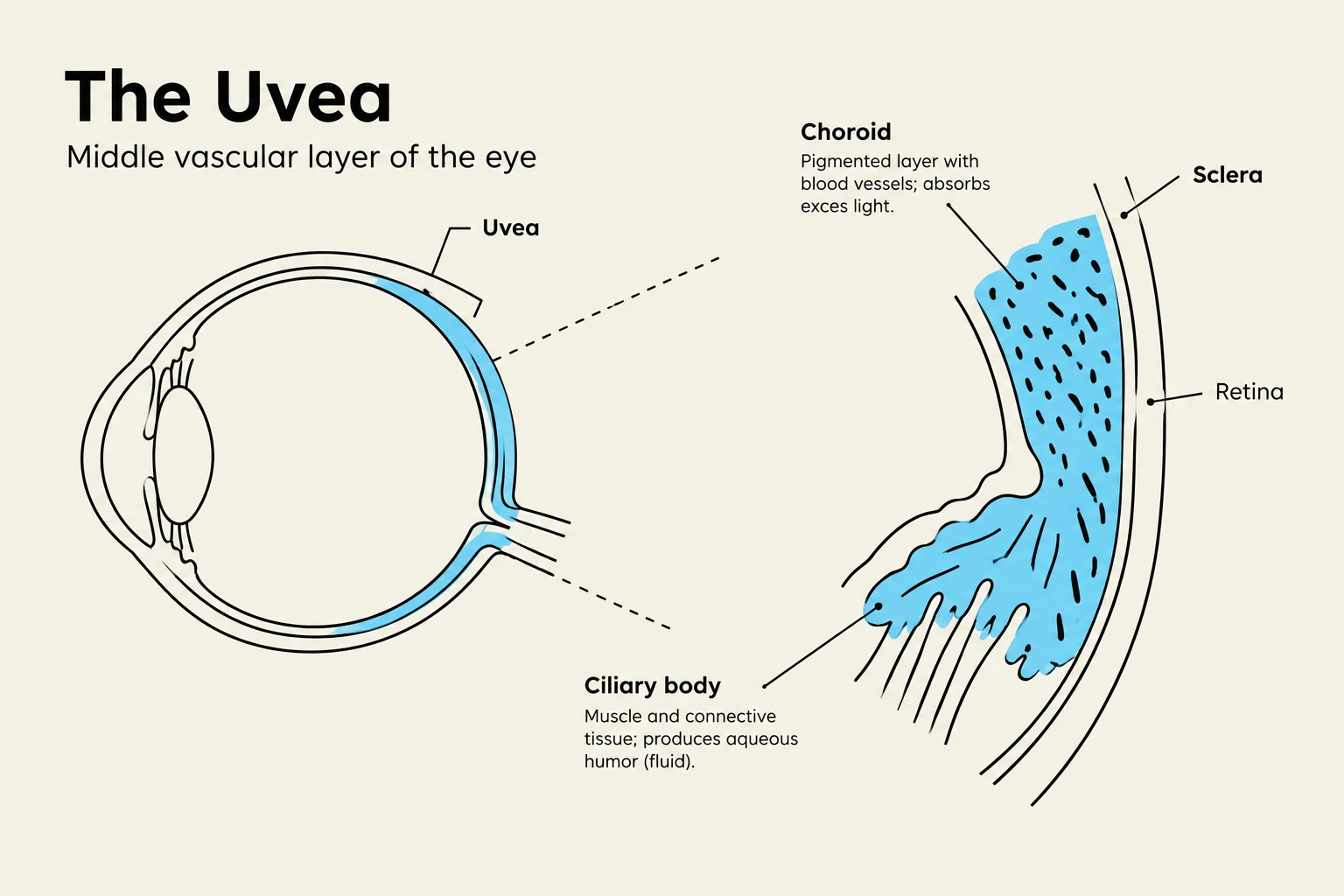
The eyeball is organized into three distinct concentric layers, often referred to as "tunics." These layers serve different primary functions: protection, nourishment, and sensory reception. Understanding the distinction between these layers is fundamental to studying ocular pathology and physiology.
1. The Fibrous Tunic (Outer Layer)
The outermost layer is the fibrous tunic, which provides structural integrity and maintains the shape of the eyeball. This layer consists of two main components: the sclera and the cornea.
- The Sclera: Often called the "white of the eye," the sclera is a tough, opaque, fibrous coat that covers most of the eyeball. It serves as an attachment point for the extrinsic muscles that move the eye.
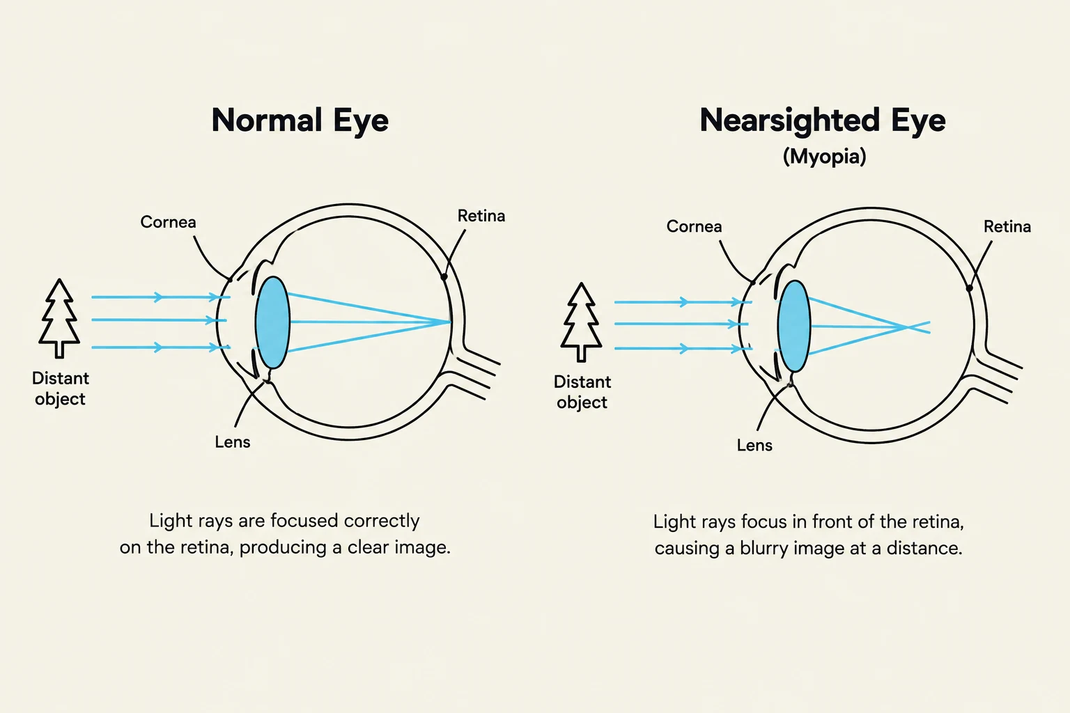
- The Cornea: At the very front of the eye, the sclera transitions into the cornea. Unlike the opaque sclera, the cornea is transparent and highly curved. It acts as the eye's primary refractive element, performing the majority of the light-bending required to focus images on the retina.

Keep reading for free.
Create an account to finish this reading and make it part of your study routine.
Continue reading freeAlready have an account? Log in
Updated