This resource explains the fundamental process of cell division, detailing the core events common to both prokaryotic and eukaryotic cells, and elaborating on the distinct phases and regulatory mechanisms of the eukaryotic cell cycle, including interphase, M phase, checkpoints, and the roles of cyclins and CDKs.
Cell division is a fundamental biological process essential for the life cycle of every organism, driving growth, regeneration, and reproduction. Whether it is a single-celled bacterium multiplying or a complex multicellular organism regenerating a lost limb, the core objective remains the same: creating new cells that carry the necessary genetic information.
While the specific mechanisms vary significantly between the simple structures of prokaryotes and the complex organelles of eukaryotes, all cell division processes must successfully execute four core events:
Cell division signals: The initial triggers that tell a cell it is time to divide.
DNA replication: The duplication of genetic material so that each daughter cell receives a complete set of instructions.
DNA segregation: The accurate distribution of the replicated DNA to the two new cells.
Cytokinesis: The physical division of the cytoplasm to form two distinct cells.
Prokaryotic Cell Division: Binary Fission
Prokaryotes, such as bacteria, reproduce through a streamlined process known as binary fission. Because prokaryotes lack a nucleus, the process is much more direct than in eukaryotic cells.
The process is typically initiated by cell division signals that are often external, such as changes in nutrient concentration or environmental conditions. Once the signal is received, DNA replication begins at a specific site on the circular chromosome called the ori (origin) and proceeds until it reaches the ter (terminus).
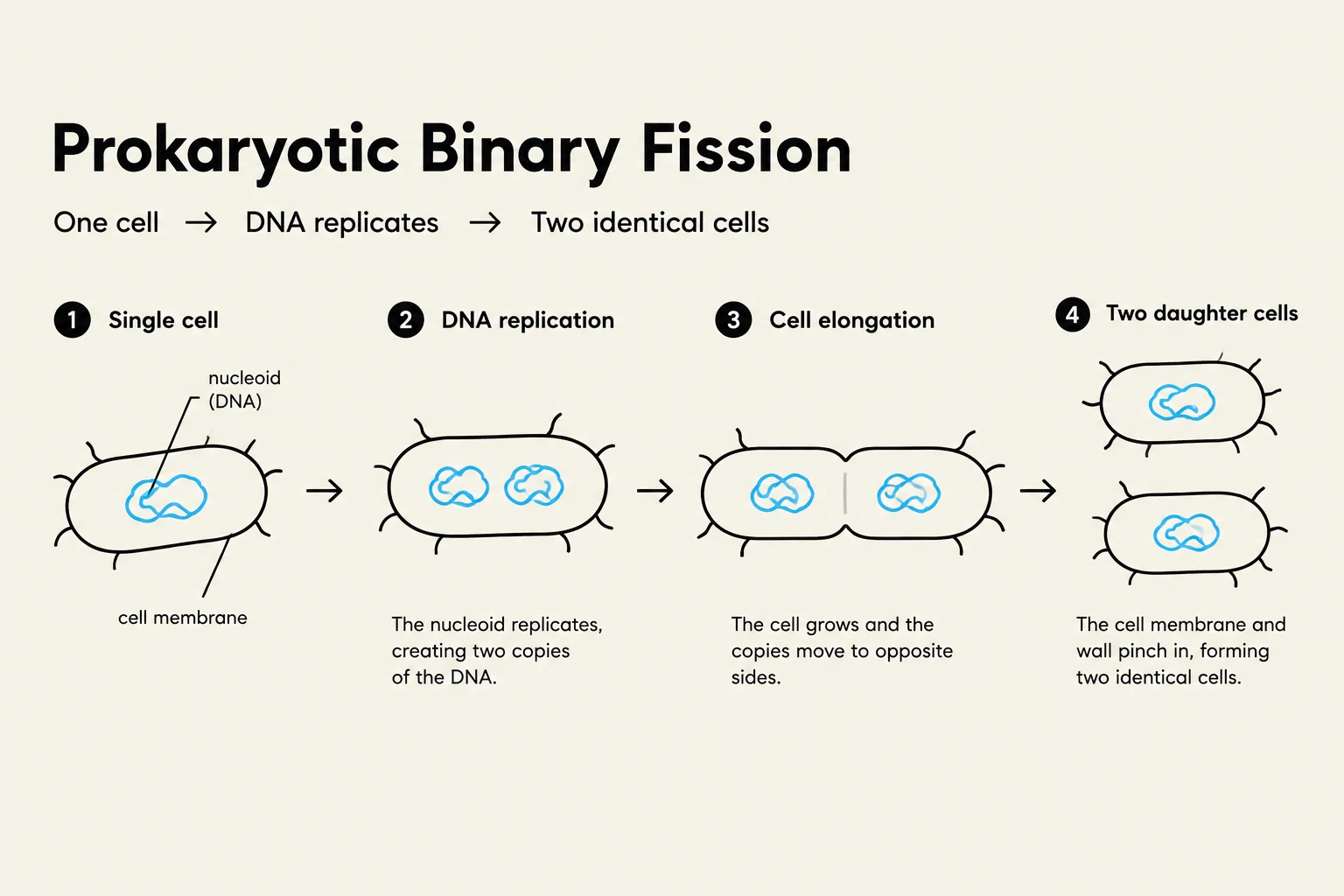
As replication occurs, the DNA moves through a protein replication complex. Following replication, DNA segregation occurs as the ori regions move toward opposite ends of the cell. Finally, cytokinesis is completed when the cell membrane pinches inward, aided by the formation of a ring of protein fibers, splitting the parent cell into two identical progeny.
Prokaryotic Binary Fission is a process where one cell divides into two identical daughter cells. This involves DNA replication, cell elongation, and finally, the pinching in of the cell membrane and wall to form two separate cells.
The Eukaryotic Cell Cycle
Eukaryotic cell division is significantly more complex due to the presence of a nucleus and multiple linear chromosomes. The eukaryotic cell cycle is broadly divided into two main stages: Interphase and the M phase.
Interphase: Preparation and Growth
Interphase is the period during which the nucleus is visible and the cell performs its normal biological functions. It is not a "resting" period, but rather a highly active phase of preparation consisting of three subphases:
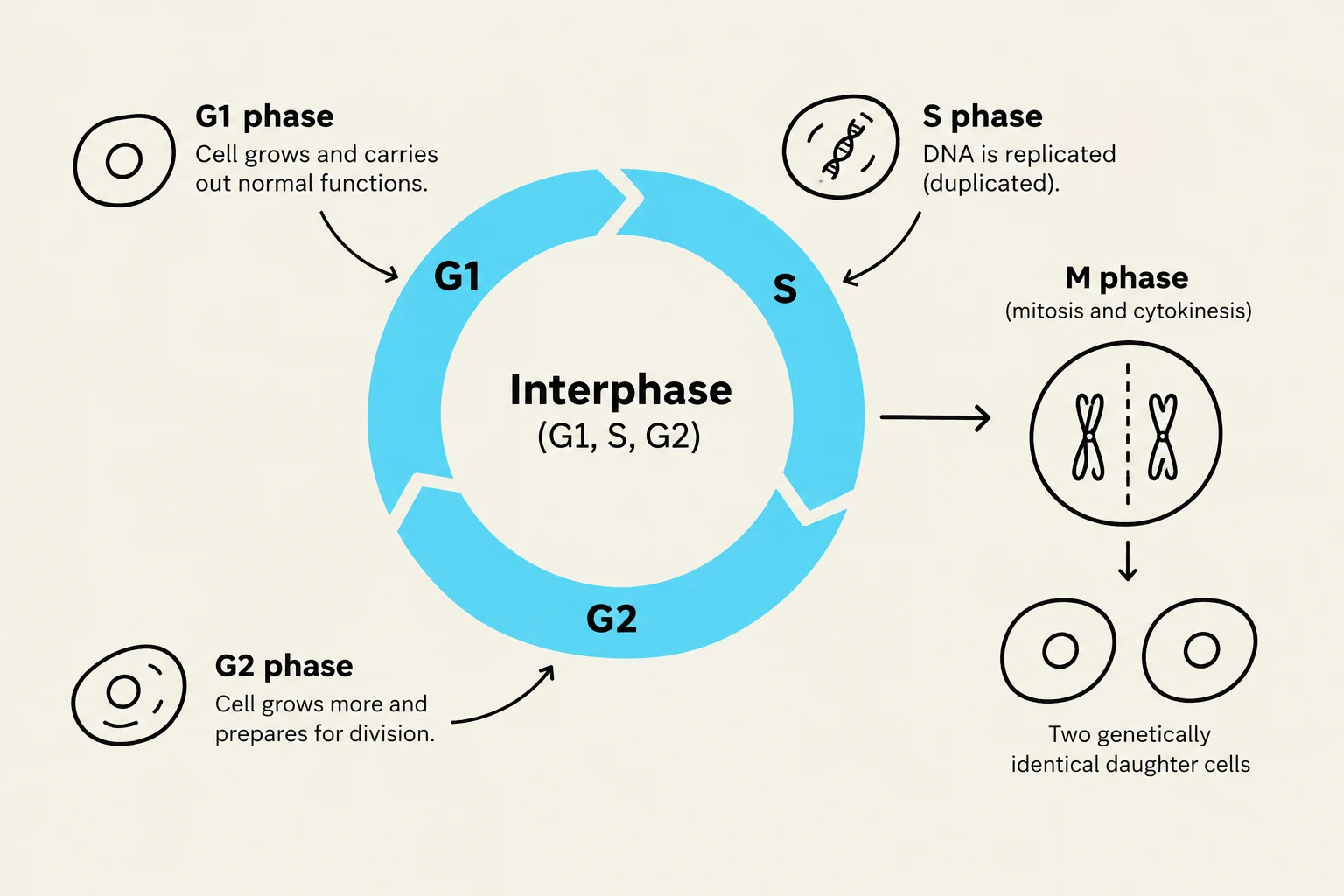
G1 phase (Gap 1): The cell grows and carries out metabolic functions. During this stage, each chromosome exists as a single, unreplicated DNA molecule. The duration of G1 can vary from minutes to years. Some cells may exit the cycle entirely at this stage to enter a non-dividing resting state called G0. The phase concludes at the G1-to-S transition, a critical point where the cell commits to DNA replication.
S phase (Synthesis): This is the stage of DNA replication. Each chromosome is duplicated to produce two identical copies known as sister chromatids. These sister chromatids remain physically joined together until they are separated during mitosis.
G2 phase (Gap 2): After replication is complete, the cell enters G2 to prepare for the upcoming division. During this phase, the cell synthesizes the specific proteins and structures required to move the chromatids to opposite ends of the cell.
The cell cycle illustrates the stages of cell division: G1 phase (growth), S phase (DNA replication), G2 phase (preparation for division), and M phase (mitosis and cytokinesis), resulting in two genetically identical daughter cells.
M Phase: Division
The M phase is the actual process of division, consisting of two distinct but coordinated events:
Mitosis: The process of nuclear division, where the replicated chromosomes are segregated into two new nuclei.
Cytokinesis: The physical division of the cytoplasm, resulting in the formation of two separate daughter cells.
Regulation: Checkpoints and Molecular Control
Because errors in DNA replication or segregation can be catastrophic (such as causing cancer), the eukaryotic cell cycle is strictly regulated by cell cycle checkpoints. These checkpoints act as control mechanisms that monitor specific conditions—such as DNA damage, nutrient availability, or whether chromosomes are properly attached to the mitotic spindle—before allowing the cell to proceed to the next phase.
Cyclin-CDK Complexes
The "engine" driving the cell cycle consists of cyclin-dependent kinases (CDKs). CDKs are enzymes (specifically kinases) that catalyze the transfer of a phosphate group from ATP to a substrate protein. However, CDKs are inactive on their own; they only become active when bound to a regulatory protein called a cyclin.
Different cyclin-CDK complexes are active at different stages of the cycle. For example, the G1/S cyclin is produced during the G1 phase, binds to its partner CDK to activate it, and is subsequently broken down to inactivate the complex and allow the cycle to progress.
The Restriction Point and RB Protein
A critical checkpoint occurs during the G1 phase, known as the restriction point (R). This point determines whether a cell will commit to the S phase and subsequent division.
The control of this point relies on the phosphorylation of RB (retinoblastoma protein):
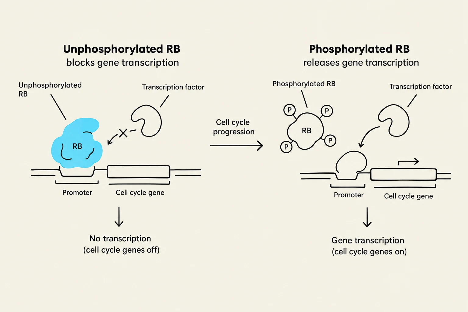
When RB is unphosphorylated: It is active and binds to transcription factors, inhibiting the expression of genes needed for DNA replication. This prevents the cell from passing the restriction point.
When RB is phosphorylated: The G1/S cyclin-CDK complex adds phosphate groups to RB. This inactivates RB, causing it to release the transcription factors. The genes for DNA replication are then expressed, and the cell moves past the restriction point into the S phase.
Exam Tip: Clinical Connection
In many types of cancer, the RB protein is mutated or becomes permanently inactive. If RB cannot function, it cannot inhibit gene transcription, meaning the cell loses its ability to stop the cell cycle at the restriction point, leading to uncontrolled cell division.
This diagram illustrates how the phosphorylation state of the RB protein affects gene transcription. Unphosphorylated RB binds to the promoter region, blocking transcription of cell cycle genes. Upon phosphorylation, RB releases the promoter, allowing transcription factors to bind and initiate gene transcription, thus promoting cell cycle progression.
Finally, it is important to note that cell division is not controlled solely by internal mechanisms. External signals, such as growth factors, can trigger signal transduction pathways that lead to the synthesis of cyclins, thereby activating CDKs and initiating the cell cycle.
Keep reading for free.
Create an account to finish this reading and make it part of your study routine.