


In the study of neurobiology, researchers often seek the perfect balance between complexity and simplicity. While the human brain is an incredibly intricate "woven feltwork" of cells, its sheer scale makes it difficult to map every single connection. Enter Caenorhabditis elegans (C. elegans), a tiny, transparent nematode worm that serves as a foundational model for understanding how nervous systems function. Despite being only about 1mm long, this organism possesses a nervous system so well-defined that scientists have mapped its entire architecture, a feat known as creating a connectome.
The Anatomy of a Nematode
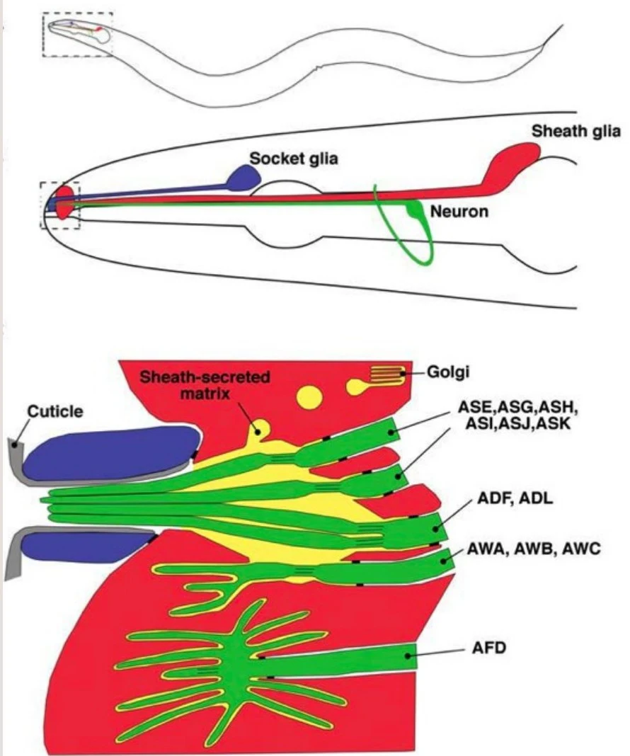
The C. elegans nervous system is remarkably efficient. The worm consists of approximately 954 cells, of which 302 are neurons and 56 are glial cells. Because the worm is transparent, researchers can use specialized techniques, such as expressing Green Fluorescent Protein (GFP), to visualize the nervous system in living specimens.

While the worm lacks a cardiovascular or respiratory system, its nervous system is highly organized. Most neurons are grouped into ganglia, and most possess long processes—axons and dendrites—that extend far beyond the cell body.
The Neuronal Trio: Sensory, Inter, and Motor Neurons
The 302 neurons in C. elegans are not all the same; they are categorized into three primary functional types that allow the worm to interact with its environment:
- Sensory Neurons: These act as the "input" devices. They detect environmental stimuli such as touch (mechanosensory), smell, taste, and temperature.
- Inter-neurons: These serve as the "processors." They act as bridges, receiving signals from sensory neurons and relaying them to the motor neurons.
- Motor Neurons: These are the "output" devices. They control muscle action, either by connecting directly to muscles at neuromuscular junctions or by signaling other neurons.
Keep reading for free.
Create an account to finish this reading and make it part of your study routine.
Continue reading freeAlready have an account? Log in
Updated