The Clotting Cascade: Hemostasis and Coagulation Pathways
An in-depth explanation of the clotting cascade, detailing the extrinsic, intrinsic, and common pathways involved in hemostasis, and the roles of key factors in converting fibrinogen to fibrin to form a stable blood clot.
When a blood vessel is damaged, the body must transition from a liquid state to a gel-like state to prevent excessive blood loss. This process, known as hemostasis, occurs in two main stages: primary hemostasis and secondary hemostasis. During primary hemostasis, blood vessels constrict and platelets aggregate to form a temporary platelet plug. However, this plug is relatively weak and requires reinforcement. This reinforcement is achieved through secondary hemostasis, more commonly known as the clotting cascade.
The clotting cascade is a complex series of enzymatic reactions where various "factors" (proteins) activate one another in a sequence. The ultimate goal of this cascade is to convert the soluble protein fibrinogen into insoluble fibrin strands, which weave together to form a stable fibrin mesh.
The Three Pathways of Coagulation
The cascade is traditionally divided into three distinct pathways: the extrinsic pathway, the intrinsic pathway, and the common pathway. While they are often studied separately, they function together to amplify the signal and produce a massive amount of fibrin quickly.
1. The Extrinsic Pathway (Tissue Factor Pathway)
The extrinsic pathway is the "fast response" system, typically initiating within 15 to 30 seconds of an injury. It is triggered by external trauma that exposes the blood to substances outside the vascular system.
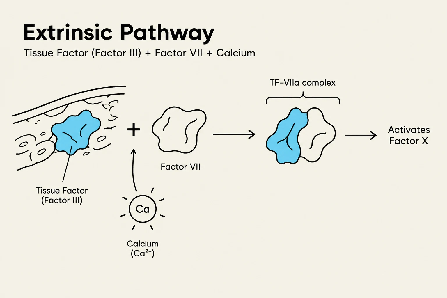
When the vasculature is damaged, Tissue Factor (Factor III) is exposed on the membranes of the damaged tissue. This Tissue Factor then encounters Factor VII circulating in the blood, activating it into Factor VIIa. In the presence of ionized calcium (Factor IV), this complex converts Factor X into Factor Xa, which officially moves the process into the common pathway.
Diagram illustrating the extrinsic pathway of blood coagulation, involving Tissue Factor (Factor III), Factor VII, and Calcium, leading to the activation of Factor X.
2. The Intrinsic Pathway (Contact Activation Pathway)
The intrinsic pathway is a slower, secondary response, often referred to as the "amplification phase". It can take anywhere from 1 to 6 minutes to fully realize. Unlike the extrinsic pathway, all the components required for this pathway are already contained within the blood itself.
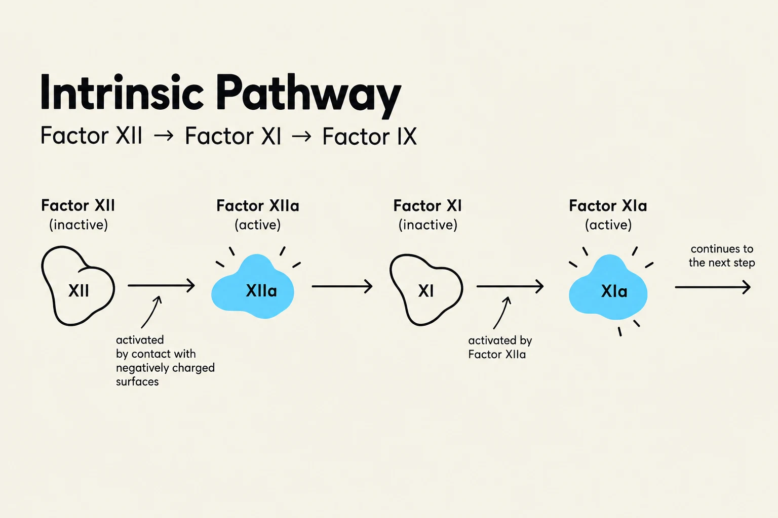
This pathway is often triggered by internal damage, such as when blood comes into contact with exposed collagen in the subendothelial layer. The sequence follows these steps:
Factor XII is activated and subsequently converts Factor XI to Factor XIa.
Factor XIa, working with calcium, activates Factor IX to Factor IXa.
Factor IXa then activates Factor VIII.
The resulting complex of Factor VIIIa, phospholipids (from platelets), and calcium activates Factor X into Factor Xa.
Diagram illustrating the intrinsic pathway of blood coagulation, showing the sequential activation of clotting factors XII, XI, and IX.
Exam Tip: Remember that the "Intrinsic" pathway is "In" the blood (all factors are circulating), whereas the "Extrinsic" pathway requires "External" tissue factor.
The Common Pathway: Creating the Mesh
Both the extrinsic and intrinsic pathways converge at a single point: the activation of Factor X (Factor 10). Once Factor Xa is produced, the common pathway begins.
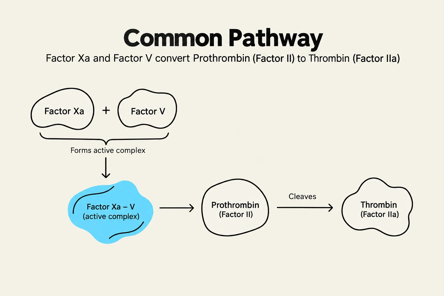
The primary objective here is the conversion of fibrinogen (Factor I) into fibrin. This is driven by thrombin (Factor IIa). Thrombin is produced via the "prothrombin activator complex," which consists of Factor Xa, Factor V, and calcium.
Once thrombin is present, it performs several critical roles:
It converts fibrinogen into fibrin monomers.
It activates Factor XIII (Fibrin Stabilizing Factor) into activated Factor 13.
It provides positive feedback by activating more Factor VII, Factor IX, and Factor V, which accelerates the entire cascade.
The final step is the most crucial for clot durability. While fibrin strands provide the structure, they are initially weak. Activated Factor 13 creates cross-links between these strands, transforming them into a strong, interconnected fibrin mesh that secures the platelet plug.
This diagram illustrates the common pathway of blood coagulation, where Factor Xa and Factor V form an active complex that cleaves Prothrombin (Factor II) into Thrombin (Factor IIa).
Key Components and Clinical Correlations
To understand the cascade in a clinical setting, such as an ICU, it is helpful to recognize the recurring "players" and how they might be affected by disease or medication.
The Role of Ionized Calcium (Factor IV)
Ionized calcium is a vital cofactor in almost every step of the cascade. Without it, the factors cannot effectively interact to activate one another. In clinical practice, it is important to distinguish between total calcium and ionized calcium, as only the ionized form is biologically available for these reactions.
Clinical Testing: PT and APTT
When clinicians need to check how well a patient's clotting cascade is functioning, they use two primary tests:
Prothrombin Time (PT/INR): This test specifically evaluates the extrinsic pathway.
Activated Partial Thromboplastin Time (APTT): This test evaluates the intrinsic pathway.
Summary Table of Factors
Factor Number
Name
Role in Cascade
I
Fibrinogen
Precursor to fibrin strands
II
Prothrombin
Precursor to Thrombin
III
Tissue Factor
Initiator of the extrinsic pathway
IV
Calcium
Essential cofactor for many steps
VII
Factor VII
Activated by Tissue Factor
VIII
Factor VIII
Cofactor in the intrinsic pathway
IX
Factor IX
Activated by Factor XIa
X
Factor X
The convergence point of all pathways
XIII
Fibrin Stabilizing Factor
Creates cross-links for the fibrin mesh
Clinical Note: Many clotting factors (specifically II, VII, IX, and X) are produced in the liver and require Vitamin K for maturation. Therefore, liver disease or Vitamin K deficiency can significantly impair a patient's ability to form a stable clot.
Keep reading for free.
Create an account to finish this reading and make it part of your study routine.