The Central Nervous System and Brain: Structure and Function
An overview of the central nervous system, detailing the organization of neural tissue, major brain regions (cerebral hemispheres, diencephalon, brain stem, cerebellum), ventricles, cerebrospinal fluid, and protective layers of the CNS.
The Central Nervous System (CNS) serves as the command center for the entire human body. It is composed of two primary structures: the brain and the spinal cord. While the spinal cord acts as a two-way conduction pathway and a center for reflexes, the brain is responsible for the most complex neural functions, including intelligence, memory, consciousness, emotion, and behavior. Furthermore, the brain regulates vital autonomic functions—such as heart rate, blood pressure, and respiration—and manages the endocrine system.
Organization of Neural Tissue
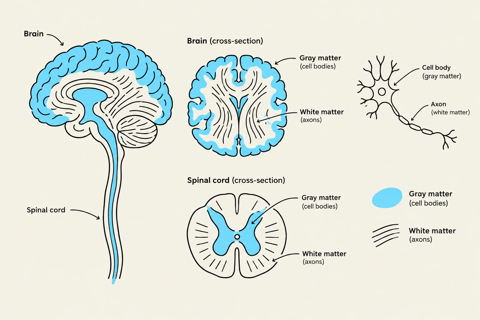
Before diving into specific regions, it is essential to understand how neural tissue is organized. The CNS is divided into gray matter and white matter.
Gray matter consists primarily of neuronal cell bodies.
White matter is composed of axons, which are often myelinated to increase the speed of signal transmission.
In the cerebrum and the cerebellum, the gray matter forms an outer layer known as the cortex.
Diagrams illustrating the structure of the brain and spinal cord, including cross-sections and a neuron.
Major Regions of the Brain
The adult brain is organized into four primary regions:
Cerebral hemispheres: The seat of high-level cognitive functions.
Diencephalon: The central core of the forebrain.
Brain stem: The passageway for fiber tracts and the origin of many cranial nerves.
Cerebellum: The center for motor coordination and equilibrium.
The Cerebral Hemispheres
The cerebral hemispheres account for approximately 83% of the total brain mass. They are divided into left and right halves by the longitudinal fissure. Each hemisphere is further subdivided into five lobes: the frontal, parietal, temporal, occipital, and insula.
The cerebral cortex is the site of the conscious mind, including sensory perception, voluntary motor initiation, and memory storage. It is highly efficient due to the presence of gyri (ridges) and sulci (grooves), which expand the surface area of the cortex.
Exam Tip: Remember that the cerebral hemispheres act contralaterally. This means the left hemisphere controls the right side of the body, and vice versa.
The Diencephalon
Located at the center of the forebrain and surrounded by the cerebral hemispheres, the diencephalon consists of three main structures:
Thalamus: Acts as the "gateway" or relay station. Almost all sensory information (except smell) must pass through the thalamic nuclei to be sorted and sent to the cerebral cortex.
Hypothalamus: The main visceral control center. It regulates the autonomic nervous system (ANS), body temperature, hunger, thirst, and the endocrine system via the pituitary gland.
Epithalamus: Includes the pineal gland, which secretes melatonin to help regulate circadian rhythms.
The Brain Stem
The brain stem serves as a critical passageway for fiber tracts connecting the cerebrum to the spinal cord. It is also the origin for 10 of the 12 pairs of cranial nerves. It is divided into three parts:
Midbrain: Contains the cerebral aqueduct and the cerebral peduncles. It houses the corpora quadrigemina, which includes the superior colliculi (visual reflexes) and inferior colliculi (auditory reflexes). Additionally, the substantia nigra (linked to Parkinson’s disease) and the red nucleus are located here.
Pons: Acts as a "bridge" connecting the cerebral cortex to the cerebellum. It is the origin for cranial nerves V (trigeminal), VI (abducens), and VII (facial).
Medulla Oblongata: Joins the spinal cord at the foramen magnum. It contains the reticular formation, which houses vital visceral centers: the cardiac center (heart rate), vasomotor center (blood pressure), and medullary respiratory centers (breathing rhythm).
The Cerebellum
Located dorsal to the pons and medulla, the cerebellum is responsible for smoothing and coordinating voluntary movements and maintaining posture and equilibrium. These processes are largely unconscious. The cerebellum receives input from the motor cortex, compares intended movement with actual body position, and sends corrective instructions back to the brain to fine-tune motor commands.
Ventricles and Cerebrospinal Fluid (CSF)
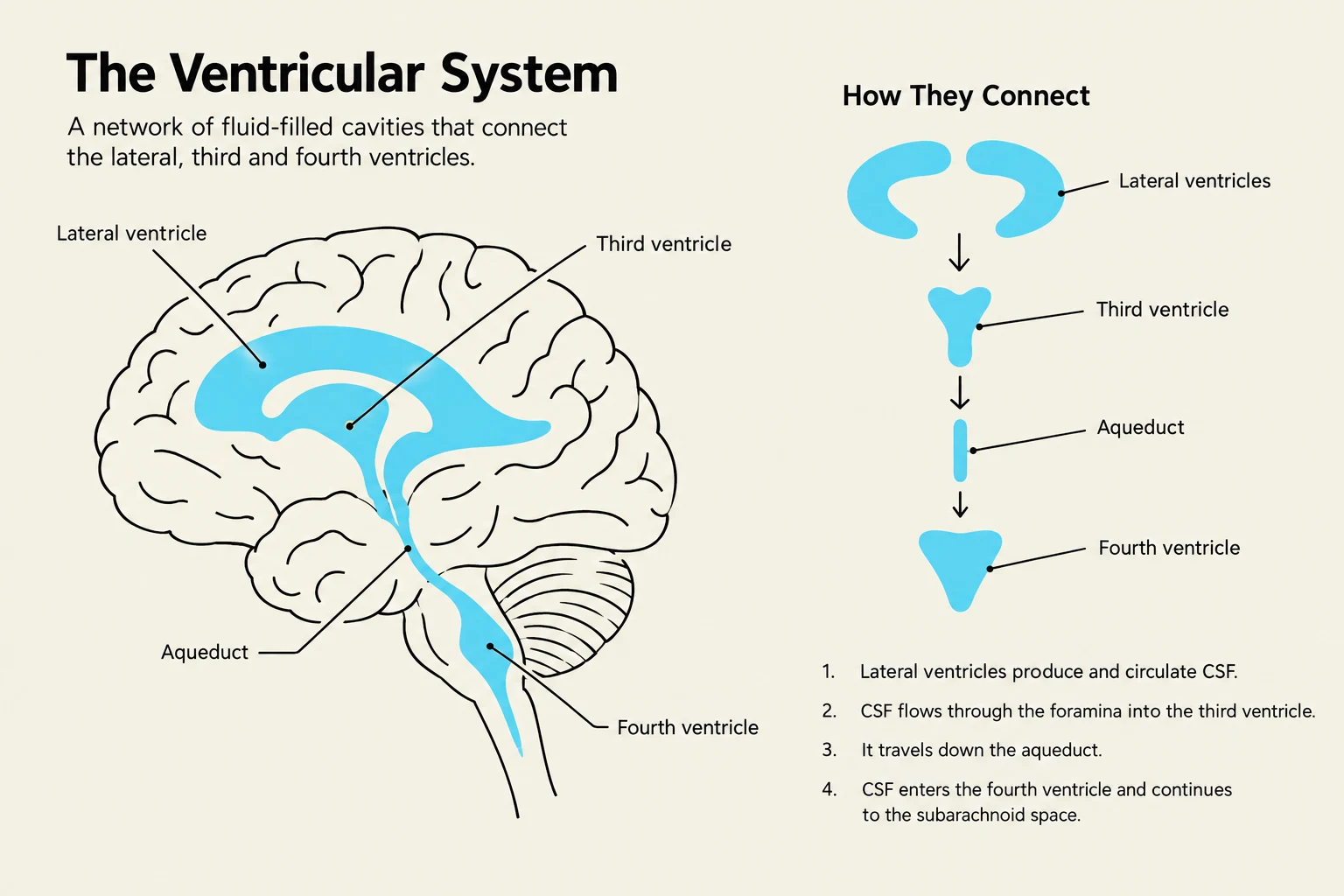
The brain is not a solid mass; it contains hollow cavities called ventricles. These ventricles are continuous with each other and the central canal of the spinal cord. They are lined with ependymal cells and filled with cerebrospinal fluid (CSF).
The ventricular system includes:
Lateral ventricles: One located in each cerebral hemisphere.
Third ventricle: Located within the diencephalon.
Fourth ventricle: Situated between the brain stem and the cerebellum.
CSF is produced in the choroid plexuses located within the ventricles. It serves several vital roles: providing a liquid cushion, nourishing the CNS, removing wastes, and carrying chemical signals.
The ventricular system of the brain, illustrating the lateral, third, and fourth ventricles and the aqueduct, along with a diagram showing the flow of cerebrospinal fluid (CSF).
Protection of the CNS
Because the CNS is so vital, it is protected by multiple layers of defense:
The Skull: Provides bony protection.
Meninges: Three protective membranes consisting of the dura mater (the strongest, outermost layer), the arachnoid mater, and the pia mater (the delicate innermost layer that clings to the brain surface).
Cerebrospinal Fluid: Provides buoyancy and cushioning.
Blood-Brain Barrier: A highly selective barrier formed by impermeable capillaries that prevents most blood-borne toxins from entering the brain, while still allowing essential nutrients like oxygen to pass.
Keep reading for free.
Create an account to finish this reading and make it part of your study routine.