The Central Dogma of Molecular Biology: DNA, RNA, and Protein Synthesis
Explains the Central Dogma of molecular biology, detailing the flow of genetic information from DNA to RNA through transcription, and from RNA to protein through translation. Covers DNA structure, nucleotides, Watson-Crick pairing, chromosomes, genes, and the roles of DNA polymerase and RNA polymerase.
Every living organism, from the simplest bacteria to the complex human body, operates based on a precise set of biological instructions. These instructions are not merely floating in space; they are organized, stored, and executed through a sophisticated molecular workflow. To understand how a cell functions, grows, and even how it fails in diseases like cancer, we must first understand the "Profile of a Cell" through its most fundamental process: the flow of genetic information.
The Central Dogma: The Blueprint of Life
At the heart of molecular biology lies the Central Dogma. This concept describes the specific, directional flow of information within a biological system: DNA → RNA → Protein.
Think of the cell like a professional kitchen. The DNA is the original, master cookbook that is far too precious to leave the safety of the office (the nucleus). To actually cook a meal, you don't take the whole book to the stove; instead, you make a photocopy of a single recipe. This photocopy is the RNA, which acts as the intermediate "middle man". Finally, the chef uses that recipe to assemble the ingredients into a finished meal: the Protein.
Exam Tip: Remember that the Central Dogma is a framework. While DNA stores the information, it is the proteins that perform the actual work, such as catalysis, defense, and movement.
The Architecture of DNA
To understand how information is copied, we must look at the structure of Deoxyribonucleic acid (DNA). DNA is a double-stranded molecule shaped like a twisted ladder, known as a double helix.
Nucleotides and Watson-Crick Pairing
The "rungs" of this ladder are made of nucleotides, which consist of a sugar, a phosphate group, and a nitrogenous base. There are four types of bases in DNA:
Adenine (A)
Thymine (T)
Cytosine (C)
Guanine (G)
The stability of the DNA molecule relies on Watson-Crick pairing. Because of their chemical shapes, certain bases always pair together: Adenine always pairs with Thymine (forming 2 hydrogen bonds), and Cytosine always pairs with Guanine (forming 3 hydrogen bonds).
Antiparallel Strands and Polymerase
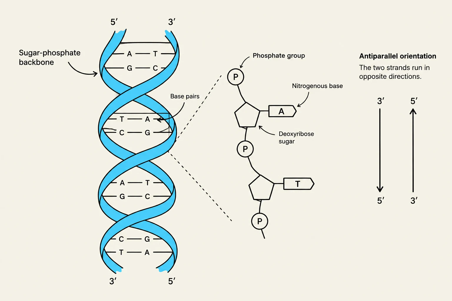
DNA strands are antiparallel, meaning they run in opposite directions. One strand runs in the 5′→3′ direction, while its complement runs 3′→5′. This orientation is determined by the carbon atoms in the sugar molecule (labeled $1'$ through $5'$).
The enzyme DNA polymerase is responsible for building new DNA strands, but it has a strict limitation: it can only attach new nucleotides to the $3'$ hydroxyl (-OH) group. Consequently, the DNA strand always grows in the 5′→3′ direction.
Diagram illustrating the structure of DNA, including the sugar-phosphate backbone, base pairs, and antiparallel orientation of the strands.
Genomic Organization: Chromosomes and Genes
DNA does not float loosely in the cell; it is tightly packaged into structures called chromosomes. This packaging keeps the massive amount of genetic information organized.
In humans, DNA is organized into 23 pairs of chromosomes, totaling 46. Humans are diploid, meaning our somatic (body) cells contain two sets of chromosomes—one inherited from each parent. These pairs, which have the same shape and size, are called homologous chromosomes.
There are two main categories of chromosomes:
Autosomes: The 22 pairs that determine all non-reproductive traits.
Sex Chromosomes: The 1 pair (XX or XY) that determines the biological sex of the individual.
Within these chromosomes lie genes, which are specific sections of DNA that code for a particular trait. However, a single gene can have different versions, known as alleles. For example, a gene for eye color might have one allele for brown eyes and another for green eyes.
From Code to Action: Transcription and Translation
The process of turning a gene into a functional protein happens in two major stages: transcription and translation.
Transcription: Making the Copy
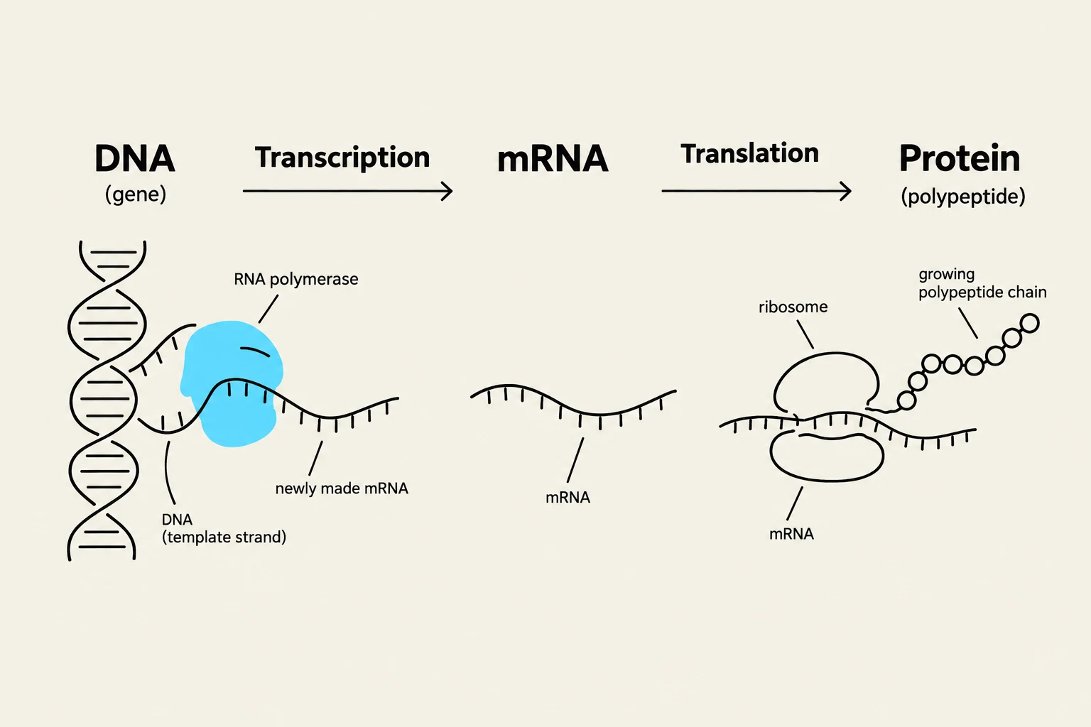
Transcription is the process where the cell uses a DNA strand as a template to create a matching RNA molecule. This is carried out by an enzyme called RNA polymerase.
A gene is not just a random string of letters; it has specific functional regions:
Promoter: The regulatory region where RNA polymerase binds to start the process.
TATA Box: A specific portion of the promoter that helps signal transcription factors to prepare the DNA for the polymerase.
Transcription Terminator: The sequence that signals the RNA polymerase to stop transcribing.
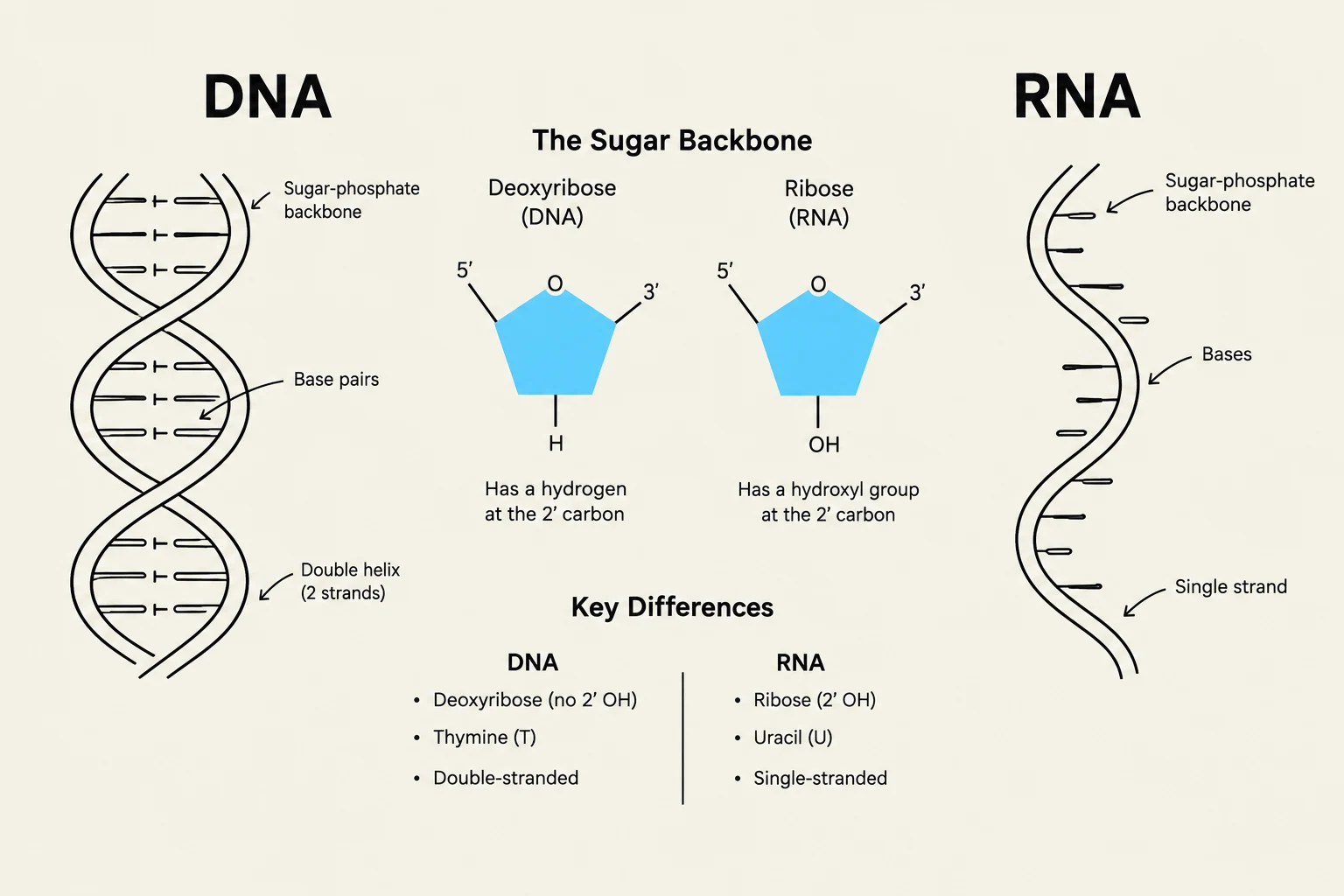
Unlike DNA, RNA uses the sugar ribose (instead of deoxyribose) and replaces Thymine (T) with Uracil (U).
Translation: Building the Protein
Once the RNA is created, the cell moves to translation. During this stage, the information in the RNA is used to assemble a chain of amino acids to form a protein.
The "language" of translation is based on triplets. A group of three bases on the RNA is called a codon. Each specific codon corresponds to one specific amino acid. Because a protein's function is entirely dependent on its shape, the specific sequence of amino acids is critical; changing even one can alter the protein's ability to work.
This diagram illustrates the central dogma of molecular biology, showing the processes of DNA transcription into mRNA and mRNA translation into a polypeptide chain (protein).
Summary Table: DNA vs. RNA
Feature
DNA
RNA
Full Name
Deoxyribonucleic acid
Ribonucleic acid
Sugar Type
Deoxyribose
Ribose
Strands
Double-stranded
Single-stranded (typically)
Nitrogenous Bases
A, T, C, G
A, U, C, G
Function
Master blueprint (storage)
Intermediate messenger
Clinical Connection: Dysregulation in these processes is a hallmark of cancer. If transcription factors or the cell cycle checkpoints fail, cells may begin to divide uncontrollably, fail to undergo apoptosis (programmed cell death), or fail to differentiate into specialized cells.
This illustration compares DNA and RNA, highlighting their structural differences, including the sugar backbone (deoxyribose vs. ribose), the presence of thymine in DNA and uracil in RNA, and their double-stranded versus single-stranded nature.
Keep reading for free.
Create an account to finish this reading and make it part of your study routine.