Explains the central dogma of molecular biology, detailing the flow of genetic information from DNA to RNA to protein, and the intricate process of DNA replication, including the roles of key enzymes and the mechanisms of leading and lagging strand synthesis.
At the heart of every living organism lies a fundamental question: how does a single molecule contain the instructions to build and operate a complex life form? The answer is found in the relationship between DNA, RNA, and proteins. This process, known as the Central Dogma, describes the unidirectional flow of genetic information from the permanent storage of DNA to the functional execution of proteins. Understanding this flow requires us to look closely at how DNA is structured, how it is copied with incredible precision, and how the molecular machinery of the cell ensures that life can persist through generations.
The Central Dogma and Genetic Information Flow
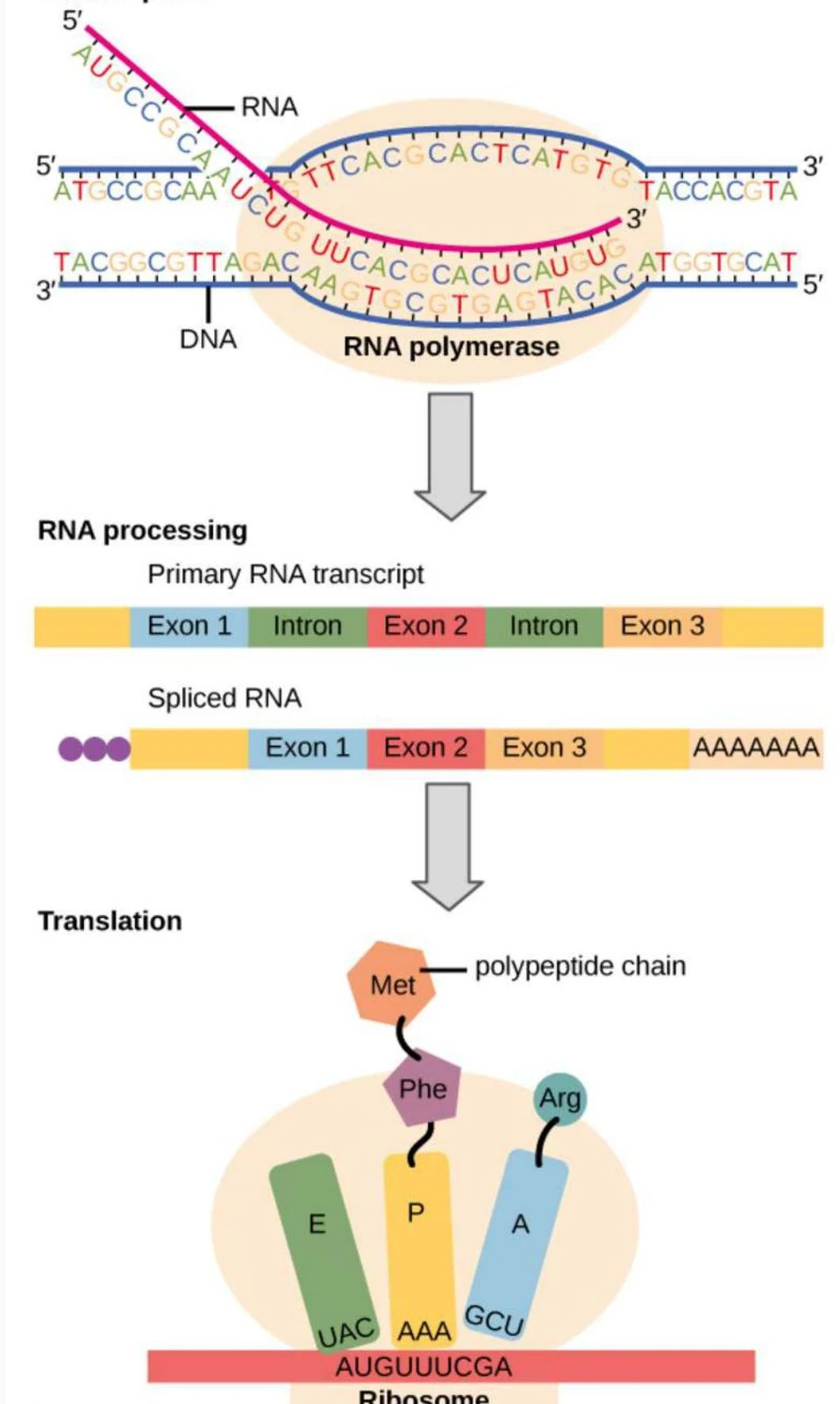
The Central Dogma serves as the roadmap for biological function. It dictates that information is stored in the sequence of nucleotides within DNA, which is then transcribed into messenger RNA (mRNA), and finally translated into a sequence of amino acids to form a protein.
This diagram illustrates the central dogma of molecular biology, showing the processes of transcription (DNA to RNA), RNA processing (intron removal and exon splicing), and translation (RNA to polypeptide chain) on a ribosome.
To understand this flow, we must distinguish between the roles of the three main players:
DNA: The long-term, stable storage of genetic information.
RNA: A short-lived, reactive messenger that transmits instructions.
Protein: The functional product that carries out cellular tasks.
The stability of DNA is due to its chemical structure. Unlike RNA, which contains a ribose sugar with a reactive hydroxyl (-OH) group at the 2' position, DNA uses deoxyribose, which lacks this oxygen atom. This small structural difference makes DNA much more chemically stable, ideal for a permanent genetic record.
The Mechanics of DNA Replication
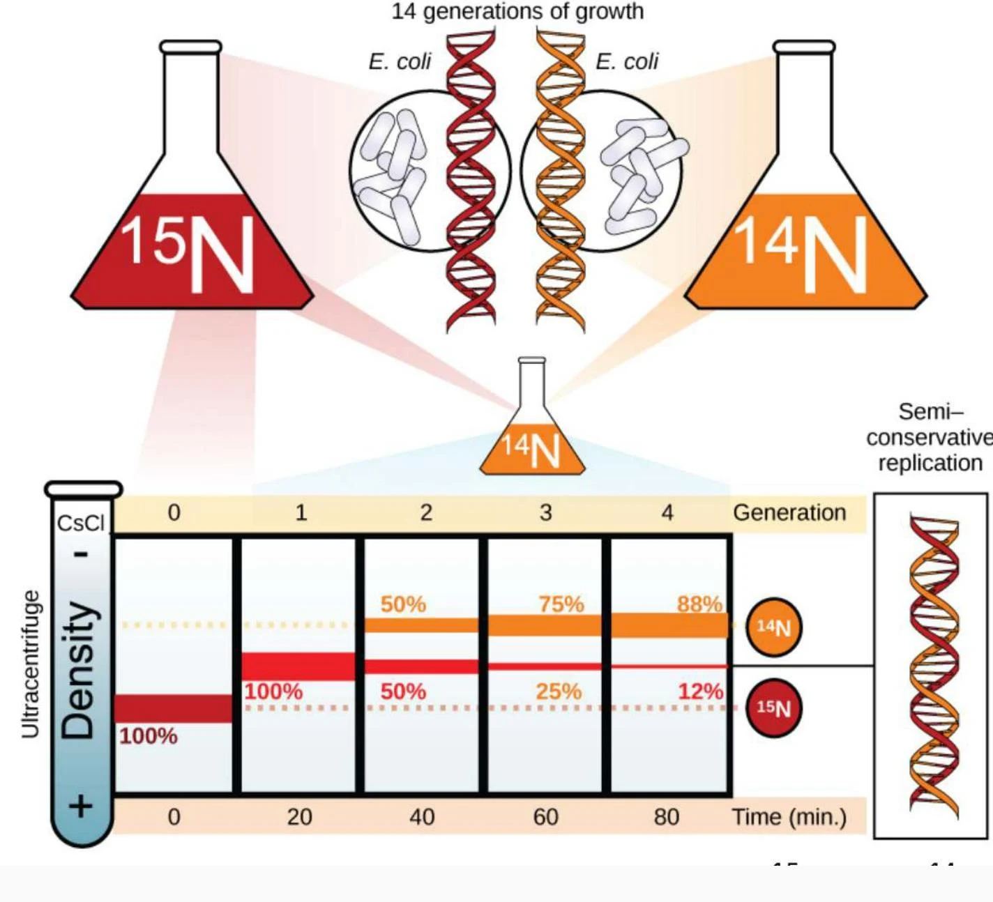
Before a cell can divide, it must create an exact copy of its entire genome. This process is called DNA replication. In 1958, Meselson and Stahl provided definitive proof that DNA replication is semi-conservative. This means that when a double helix is copied, the two original strands separate, and each serves as a template for the synthesis of a new, complementary strand.
This illustration depicts the semi-conservative replication of DNA in E. coli, showing how DNA strands separate and each serves as a template for a new strand. The experiment uses isotopes of nitrogen (15N and 14N) and ultracentrifugation to track DNA distribution across generations.
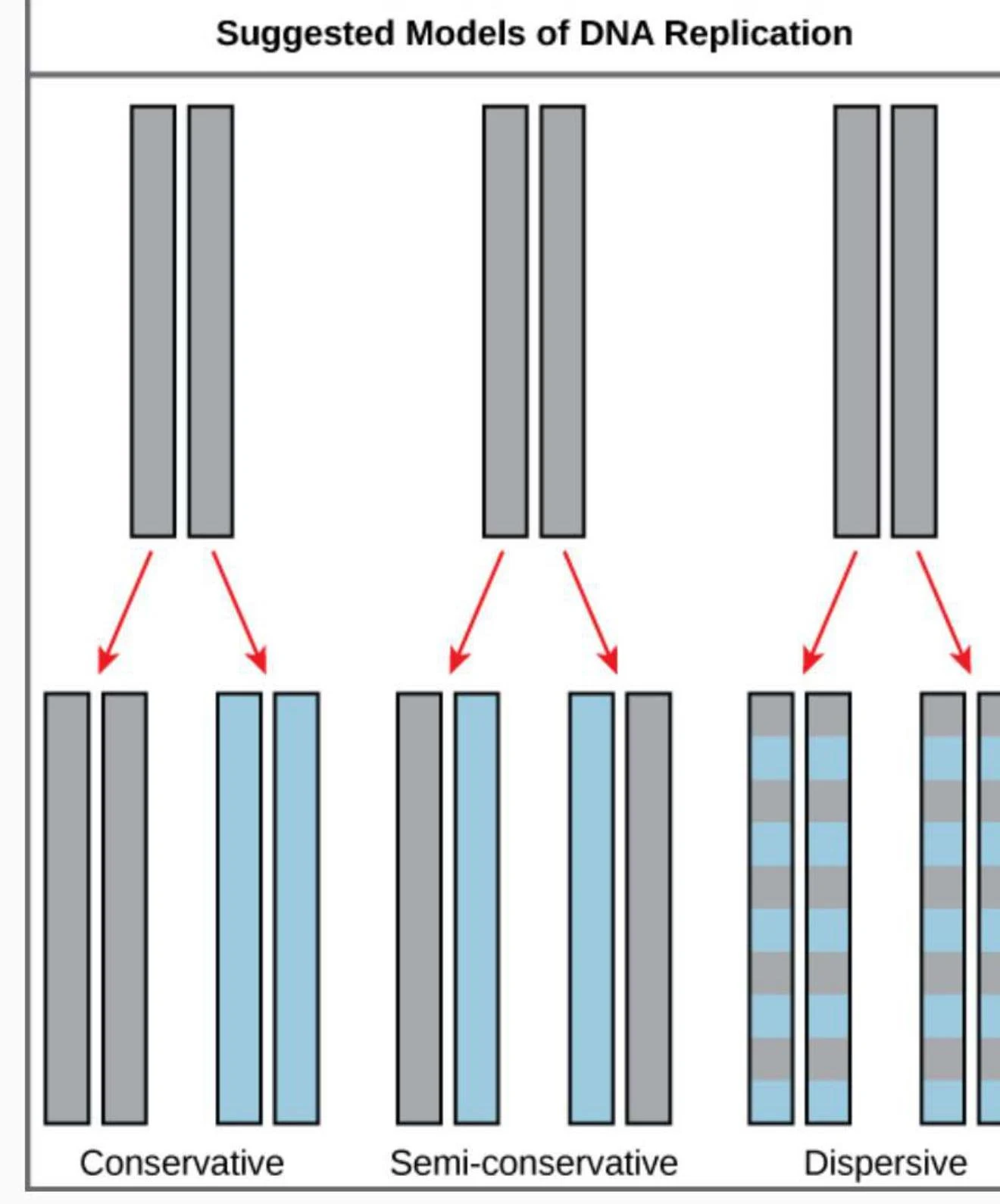
Diagrams illustrating three models of DNA replication: conservative, semi-conservative, and dispersive.
As shown in the models above, the semi-conservative method ensures that each daughter molecule consists of one "old" parental strand and one "new" synthesized strand.
The Replisome: The Molecular Machinery
Replication is not a passive process; it is driven by a complex assembly of enzymes and proteins collectively known as the replisome. The process begins at specific sites called origins of replication, where the double helix is unwound.
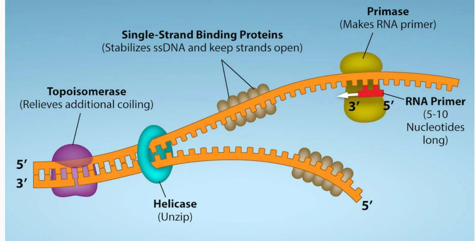
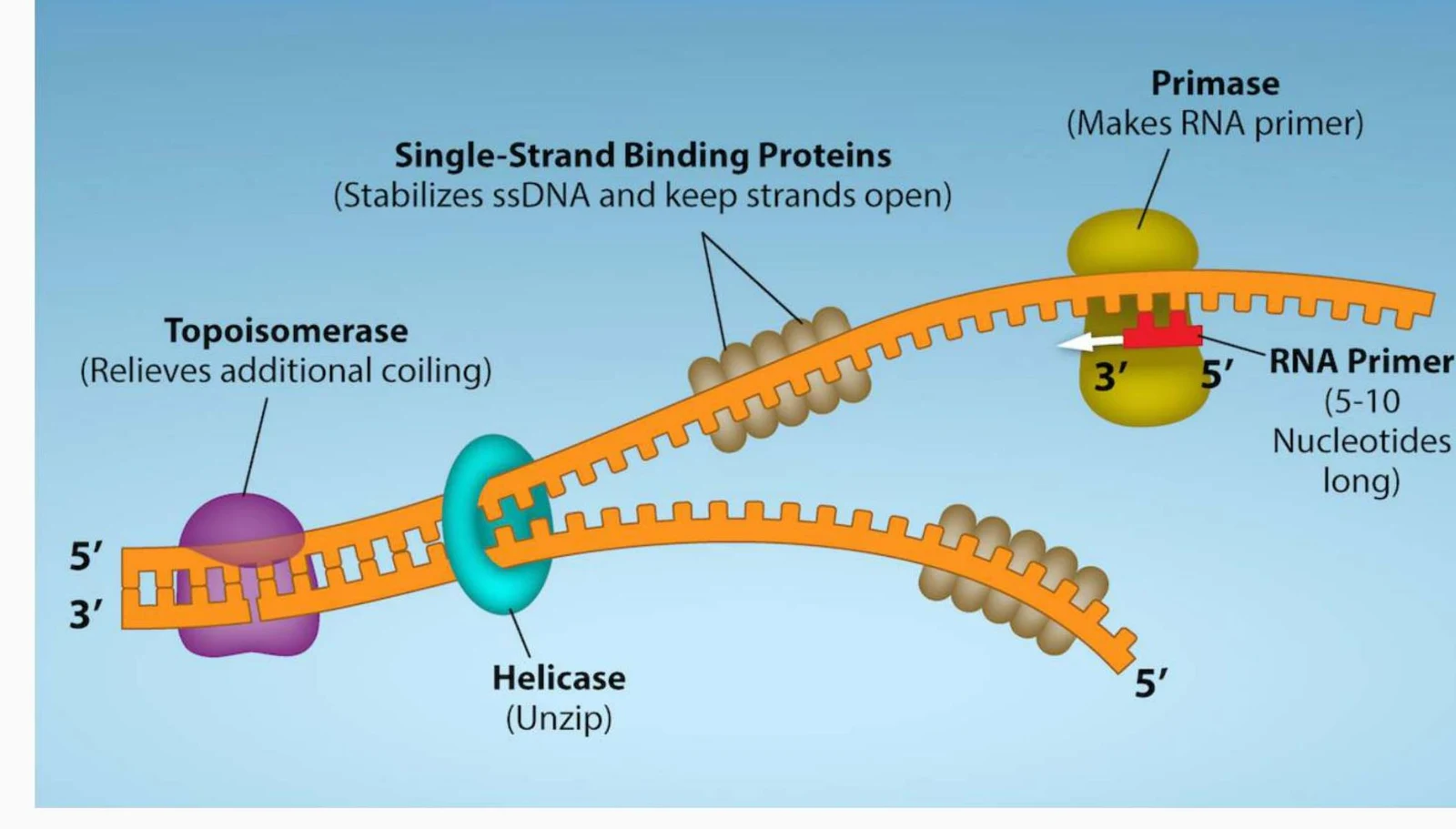
Helicase: This enzyme acts like a zipper, breaking the hydrogen bonds between the nitrogenous bases to unwind the double helix.
Topoisomerase: As helicase unwinds the DNA, the helix ahead of the fork becomes tightly coiled and stressed. Topoisomerase relieves this tension by cutting, rotating, and rejoining the DNA strands.
Primase: DNA polymerase cannot start a new strand from scratch; it can only add nucleotides to an existing chain. Primase solves this by synthesizing a short RNA primer, providing the necessary 3'-OH starting point.
DNA Polymerase: This is the primary builder. It reads the template strand and adds complementary nucleotides in a 5′→3′ direction.
Ligase: Once the RNA primers are removed and replaced with DNA, ligase acts as the "glue," sealing the gaps between fragments to create a continuous strand.
Diagram illustrating the process of DNA replication, showing the roles of Topoisomerase, Helicase, Single-Strand Binding Proteins, and Primase in unwinding and stabilizing the DNA strands, and initiating synthesis with an RNA primer.
Diagram illustrating DNA replication with key enzymes like helicase, topoisomerase, primase, and single-strand binding proteins.
Leading vs. Lagging Strands
Because DNA strands are antiparallel (running in opposite directions) and DNA polymerase can only synthesize in the 5′→3′ direction, the two strands are replicated differently:
The Leading Strand: This strand is synthesized continuously toward the replication fork.
The Lagging Strand: This strand must be synthesized in the opposite direction of the fork's movement. It is created in short, discontinuous segments called Okazaki fragments.
Exam Tip: Remember that the directionality of DNA is determined by the carbon numbering of the sugar. The $5'$ end has a phosphate group, while the $3'$ end has a hydroxyl (-OH) group. DNA polymerase always adds to the $3'$ end.
Biotechnology: Reading the Code
Our understanding of the replisome has allowed scientists to "hijack" these mechanisms for biotechnology. One of the most famous applications is Sanger sequencing.
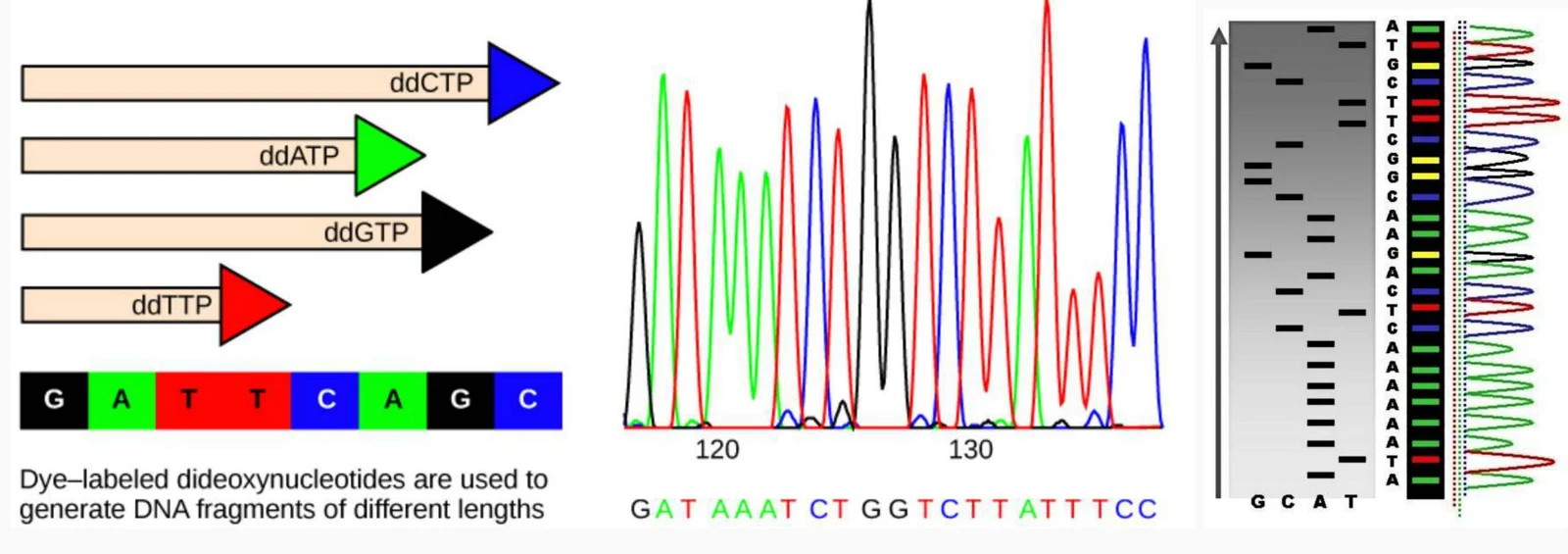
In this method, scientists use modified nucleotides called dideoxynucleotides (ddNTPs). Unlike normal nucleotides, ddNTPs lack the 3'-OH group required for the next nucleotide to attach. When a DNA polymerase incorporates a ddNTP, synthesis stops immediately. By using a collection of these "stop" nucleotides, scientists can create DNA fragments of varying lengths that reveal the exact sequence of the gene.
This illustration depicts the process of DNA sequencing using dye-labeled dideoxynucleotides, which generate DNA fragments of varying lengths. The resulting fragments are then analyzed to determine the DNA sequence, visualized here as chromatograms and a gel-like representation.
Summary of Replication Steps
To consolidate your learning, follow these ten essential steps of replication:
DNA unwinds at the origin.
Helicase opens the helix, creating two forks.
Single-strand binding proteins stabilize the strands.
Topoisomerase relieves supercoiling.
Primase lays down RNA primers.
DNA Polymerase III adds nucleotides to the $3'$ end.
Elongation occurs on the leading strand (continuous) and lagging strand (Okazaki fragments).
RNA primers are removed.
DNA Polymerase I fills the gaps with DNA.
Ligase seals the fragments together.
Keep reading for free.
Create an account to finish this reading and make it part of your study routine.