The Cardiovascular System and Heart Failure: A Clinical Overview
An overview of the cardiovascular system, including risk factors for cardiovascular disease, signs and symptoms, valvular heart disease, and congestive heart failure (CHF) categories (systolic vs. diastolic, left-sided vs. right-sided). It also covers CHF interventions, including pharmacological and lifestyle modificat
The cardiovascular system is the body's primary transport network, responsible for delivering oxygen and nutrients to tissues while removing metabolic waste for elimination. At its center is the heart, a muscular organ located in the mediastinum that functions as a pump to drive blood through pulmonary and systemic circulation. Because the cardiovascular system is a top cause of illness and death in the United States, understanding its mechanics, risk factors, and failure modes is essential for clinical practice.
Risk Factors for Cardiovascular Disease
Cardiovascular disease (CVD) does not occur in a vacuum; it is driven by a combination of factors that can be categorized into those we cannot change and those we can influence.
Non-Modifiable Risk Factors
These are inherent biological factors that a patient cannot alter. They include:
Age: Risk generally increases as a person ages.
Gender: Biological sex can influence risk profiles.
Genetics: Family history and genetic predisposition play a significant role.
Race and Ethnicity: Certain populations may have higher statistical risks.
Modifiable Risk Factors
These are lifestyle or medical conditions that can be managed or reversed through intervention:
Hypertension (High Blood Pressure): Often called the "silent killer" because it is frequently asymptomatic.
Diabetes Mellitus: High blood sugar can damage blood vessels over time.
Obesity and Physical Inactivity: Sedentary lifestyles contribute to metabolic strain.
Smoking and High Cholesterol: These directly contribute to the buildup of plaque in the arteries.
Recognizing Cardiovascular Signs and Symptoms
Clinicians must be vigilant in identifying signs of cardiovascular distress. Symptoms can range from subtle fatigue to life-threatening emergencies.
Common clinical presentations include:
Chest Pain: This may manifest as angina (chest pain after exertion) or more severe crushing pain during a myocardial infarction.
Dyspnea: Shortness of breath, which may occur during exertion or at rest.
Edema: Swelling, particularly in the ankles, legs, or sacral area.
Clinical Tip: If a patient reports sudden, crushing chest pain that lasts longer than 20 minutes and is unrelieved by rest or medication, seek immediate medical attention, as this may indicate a myocardial infarction.
Valvular Heart Disease
Diagrams illustrating the mechanisms of valvular heart disease, specifically stenosis and regurgitation.
The heart relies on four valves to ensure one-way blood flow: the atrioventricular valves (Tricuspid and Mitral) and the semilunar valves (Aortic and Pulmonary). Valvular heart disease occurs when these valves are damaged, leading to two primary mechanical issues:
Stenosis: The valve leaflets become thick, stiff, or fused, preventing the valve from opening adequately. This restricts blood flow through the valve.
Regurgitation (Insufficiency/Incompetence): The valve fails to close tightly, allowing blood to leak backward through the valve.
Symptoms of valvular disease often include palpitations, shortness of breath, angina, and fatigue.
Congestive Heart Failure (CHF)
Congestive Heart Failure occurs when the heart is unable to pump enough blood to meet the metabolic demands of the body. It is a chronic condition that is managed rather than cured. CHF is categorized by the specific nature of the pump failure.
Functional Categories: Systolic vs. Diastolic Failure
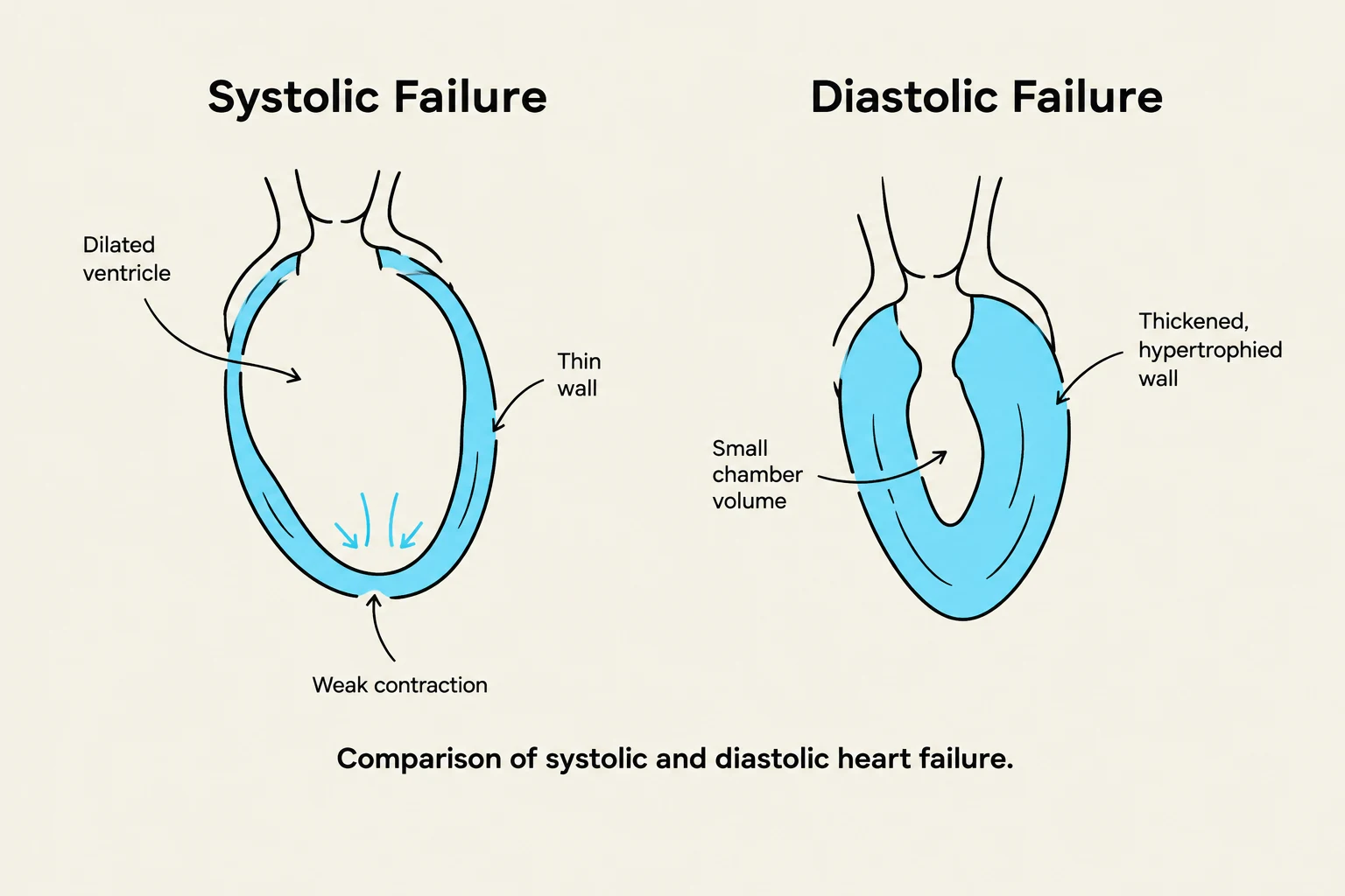
Comparison of systolic and diastolic heart failure.
Systolic Failure: A problem with the pumping mechanism. The heart cannot contract with enough force to eject an adequate amount of blood, often leading to thinner heart walls.
Diastolic Failure: A problem with filling. The heart cannot relax or expand enough to receive an adequate volume of blood, often caused by thickening of the ventricular walls or hypertension.
Anatomical Categories: Left-Sided vs. Right-Sided Failure
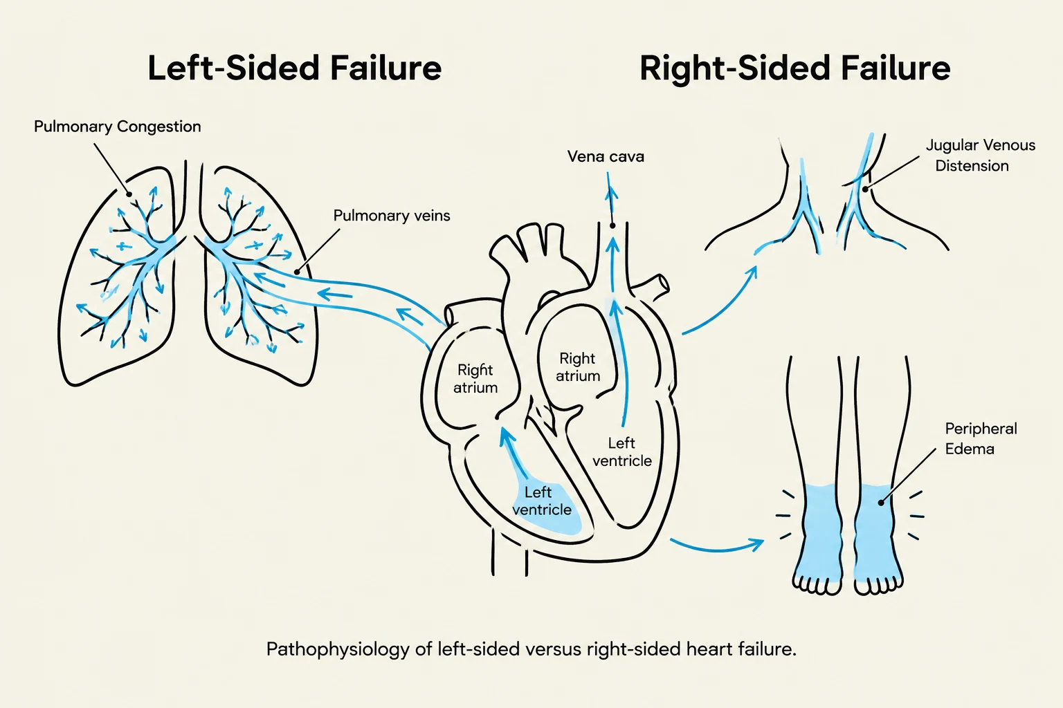
Diagram illustrating the pathophysiology of left-sided versus right-sided heart failure, showing symptoms like pulmonary congestion, jugular venous distension, and peripheral edema.
Left-Sided Heart Failure
Left-sided failure is often linked to hypertension, hyperthyroidism, or left ventricle infarction. It can also be caused by mitral valve stenosis or mitral valve prolapse. Because the left side of the heart handles oxygenated blood from the lungs, failure here causes blood to "back up" into the pulmonary veins and lungs.
Clinical signs of Left-Sided Failure include:
Pulmonary congestion (cough, crackles, wheezes, or blood-tinged sputum).
Orthopnea (difficulty breathing while lying flat) and Paroxysmal Nocturnal Dyspnea.
Tachypnea (rapid breathing), tachycardia, and cyanosis.
Right-Sided Heart Failure (Cor Pulmonale)
Right-sided failure occurs when the right side of the heart cannot efficiently pump blood into the lungs. This is often caused by pulmonary hypertension or chronic pulmonary problems like COPD (Cor Pulmonale). This failure causes blood to back up into the venous circulatory system.
Clinical signs of Right-Sided Failure include:
Dependent Edema: Pitting edema in the legs or sacral area.
Jugular Venous Distension: Visible swelling in the neck veins.
Systemic Congestion: Enlarged liver and spleen (hepatosplenomegaly), ascites (abdominal swelling), and gastrointestinal distress.
CHF Interventions and Management
Management of CHF focuses on reducing the workload of the heart and increasing its efficiency.
Pharmacological Interventions
Diuretics: Remove excess water and sodium to decrease blood pressure and reduce edema.
ACE Inhibitors: Control hypertension by inhibiting enzymes that cause vessel constriction.
Beta-Blockers: Slow the heart rate by blocking the effects of epinephrine.
Digitalis Preparations: Increase the force of cardiac contraction.
Vasodilators: Open blood vessels to reduce resistance.
Lifestyle and Surgical Interventions
Patients are encouraged to maintain a low-fat, low-sodium, and low-cholesterol diet, alongside regular exercise and weight management. In more advanced cases, surgical options such as a Cardiac Pacemaker or a Ventricular Assistive Device (VAD) may be implanted to regulate or assist the heart's pumping action.
Note for Physical Therapist Assistants (PTAs): When working with patients with cardiovascular conditions, always monitor vital signs, be aware of medication side effects (such as dizziness from beta-blockers), and educate patients on lifestyle modifications.
Keep reading for free.
Create an account to finish this reading and make it part of your study routine.