


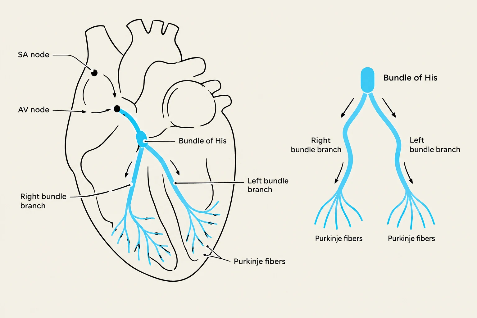
The heart is more than just a mechanical pump; it is an electrically driven organ. For the heart to pump blood effectively, the chambers must contract in a highly coordinated sequence. This coordination is made possible by the cardiac conduction system, a specialized network of nodes and fibers that generate and transmit electrical impulses throughout the myocardium. Understanding this pathway is fundamental to interpreting electrocardiograms (ECGs) and recognizing life-threatening arrhythmias.
The Pacemaker: The Sinoatrial (SA) Node
The journey of a single heartbeat begins in the right atrium at a specialized cluster of cells known as the Sinoatrial (SA) node. Often referred to as the "natural pacemaker" of the heart, the SA node is responsible for initiating the electrical impulse that sets the rhythm for the entire cardiac cycle.
Under normal physiological conditions, the SA node fires spontaneously at a rate that dictates the sinus rhythm. Once the impulse is generated, it spreads across the atria like a wave, causing the atrial muscle cells to depolarize and contract. This contraction pushes blood from the atria down into the ventricles.
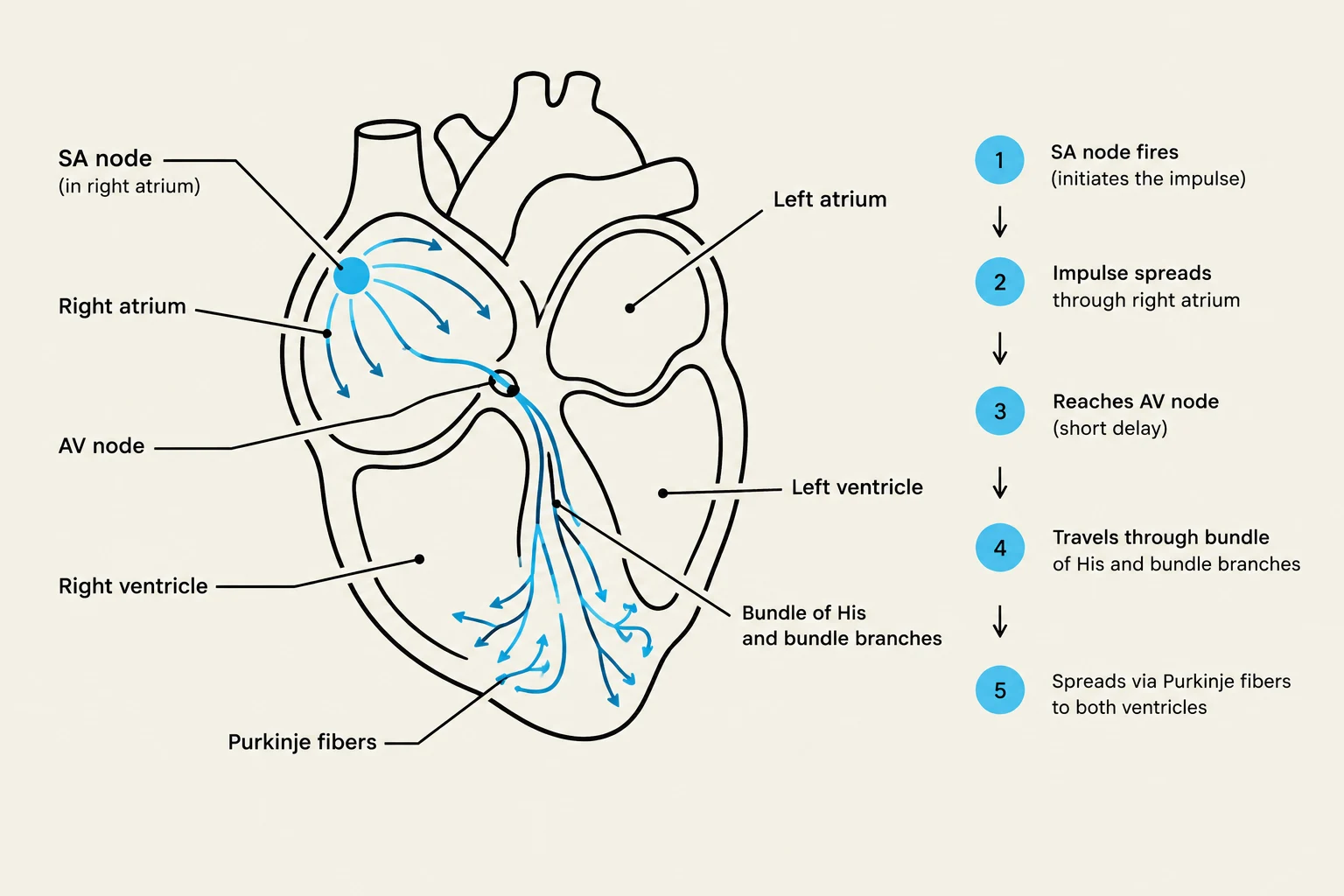
Clinical Note: If the SA node fails to function or is suppressed, other parts of the conduction system (such as the AV node) can take over as secondary pacemakers, though they typically operate at a much slower intrinsic rate.
The Gatekeeper: The Atrioventricular (AV) Node
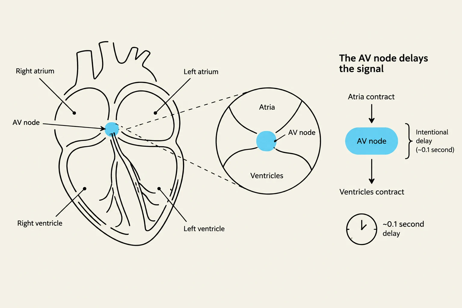
Once the electrical impulse has traveled through the atria, it reaches a critical junction: the Atrioventricular (AV) node, located near the junction of the atria and ventricles.
Keep reading for free.
Create an account to finish this reading and make it part of your study routine.
Continue reading freeAlready have an account? Log in
Updated