The Autonomic Nervous System: Structure and Function
An overview of the autonomic nervous system (ANS), its divisions (sympathetic and parasympathetic), and their roles in regulating involuntary bodily functions. Covers anatomical pathways, neurotransmitters, and integration.
The human nervous system is a complex network responsible for maintaining homeostasis—the stable internal environment required for survival. While some aspects of our behavior are conscious and controlled, much of our body's vital work happens "behind the scenes." This unconscious regulation of smooth muscle, cardiac muscle, and glands is the primary responsibility of the autonomic nervous system (ANS).
Somatic vs. Autonomic: Understanding the Difference
To understand the autonomic nervous system, it is helpful to first look at its counterpart: the somatic nervous system. The nervous system is broadly divided based on the types of functions it controls and the neurons it utilizes.
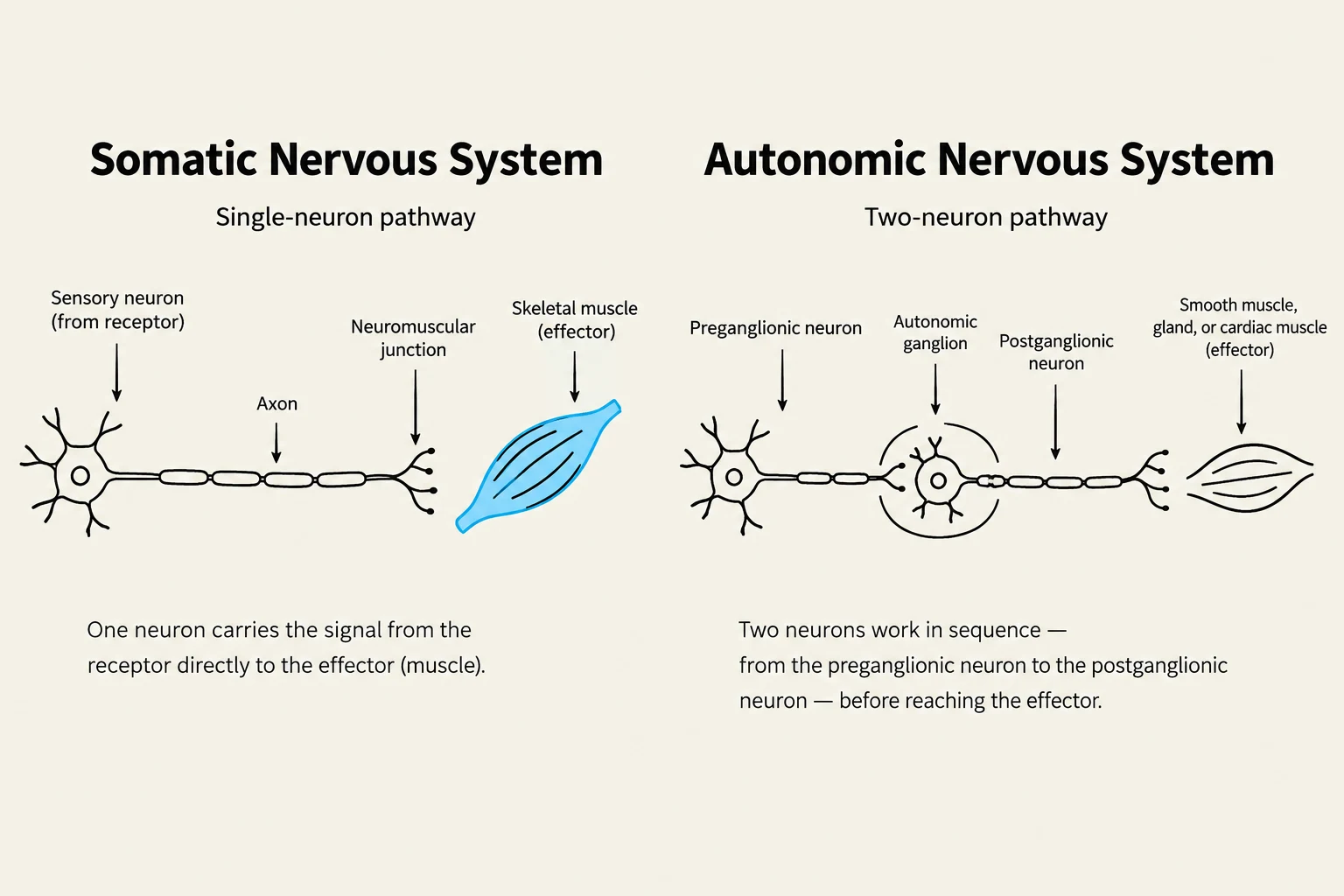
The somatic nervous system is responsible for voluntary movements and conscious sensations. It utilizes sensory neurons to relay information regarding touch, pain, temperature, sight, hearing, taste, smell, and proprioception (the sense of self-position). It also employs motor neurons that innervate skeletal muscles, allowing for intentional movement. In the somatic system, a single, myelinated motor neuron extends directly from the central nervous system (CNS) to the skeletal muscle fiber it controls.
In contrast, the autonomic nervous system manages involuntary functions. It receives input from sensory receptors located in organs, blood vessels, muscles, and the nervous system itself. Unlike the "one-neuron" pathway of the somatic system, most autonomic motor pathways consist of a chain of two neurons in series.
Diagrams illustrating the somatic nervous system (single-neuron pathway) and the autonomic nervous system (two-neuron pathway).
The Anatomy of Autonomic Pathways
The two-neuron structure of the ANS is a defining anatomical feature. These two neurons are:
The preganglionic neuron: This neuron has its cell body located within the central nervous system (the brain or spinal cord).
The postganglionic neuron: This neuron has its cell body and dendrites located in an autonomic ganglion, where it synapses with the preganglionic axon. Its unmyelinated axon then extends from the ganglion to the effector organ, such as a gland or smooth/cardiac muscle.
An autonomic ganglion serves as the essential relay station where the first neuron meets the second.
The Two Divisions: Sympathetic and Parasympathetic
The autonomic nervous system is divided into two functional branches that generally act in opposition to one another to maintain balance.
The Sympathetic Nervous System (Fight-or-Flight)
The sympathetic division is often called the "fight-or-flight" division. When stimulated, it increases alertness and metabolism, preparing the body for an emergency.
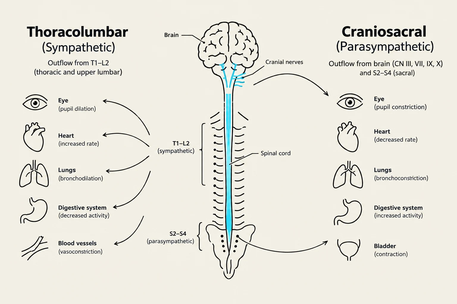
Anatomically, this is known as the thoracolumbar division because the cell bodies of the preganglionic neurons are located in the lateral horns of the gray matter in the 12 thoracic and the first 2 or 3 lumbar segments of the spinal cord. In this division, preganglionic neurons are relatively short, while postganglionic neurons are relatively long. A single sympathetic preganglionic neuron often synapses with many postganglionic neurons, allowing the signal to spread widely to various organs.
There are two major types of sympathetic ganglia:
Sympathetic trunk ganglia: These lie in a vertical row on either side of the vertebral column.
Prevertebral ganglia: These lie anterior to the vertebral column, close to the large abdominal arteries.
The Parasympathetic Nervous System (Rest-and-Digest)
The parasympathetic division is known as the "rest-and-digest" division. Its stimulation slows down most body activities, allowing the body to conserve energy and perform maintenance tasks.
This is referred to as the craniosacral division. The preganglionic cell bodies are located in the nuclei of four cranial nerves (III, VII, IX, and X) in the brainstem and in the lateral gray matter of the sacral segments 2–4 of the spinal cord.
In the parasympathetic pathway, the preganglionic neurons are long and extend a considerable distance before synapsing in terminal ganglia, which are embedded in or very near the target effectors. Consequently, the postganglionic neurons are very short and typically synapse with neurons that lead to a single, specific effector rather than a widespread area.
Diagram illustrating the effects of the sympathetic (thoracolumbar) and parasympathetic (craniosacral) nervous systems on various organs.
Neurotransmitters and Control
The way these systems communicate with organs depends on the chemicals they release, known as neurotransmitters.
Cholinergic neurons: These release the neurotransmitter acetylcholine (ACh). All preganglionic neurons (both sympathetic and parasympathetic) are cholinergic. Additionally, all parasympathetic postganglionic neurons are cholinergic.
Adrenergic neurons: These release norepinephrine (also known as noradrenalin). In the sympathetic division, the postganglionic neurons are typically adrenergic.
Exam Tip: Remember that while most sympathetic postganglionic neurons are adrenergic, sympathetic postganglionic neurons that innervate sweat glands are actually cholinergic!
The receptors that receive these chemicals determine the response. For example, nicotinic receptors always cause depolarization, while muscarinic receptors can either depolarize or hyperpolarize the membrane.
Integration and Autonomic Tone
The body does not simply switch between "on" and "off" for these systems. Instead, most organs receive dual innervation from both divisions. The balance between the two is known as autonomic tone. This tone is controlled by the hypothalamus, which acts as the major integration center. The hypothalamus receives information from various sources, including sensory input, temperature, osmolarity, and the limbic system (which handles emotions), to adjust the autonomic output accordingly.
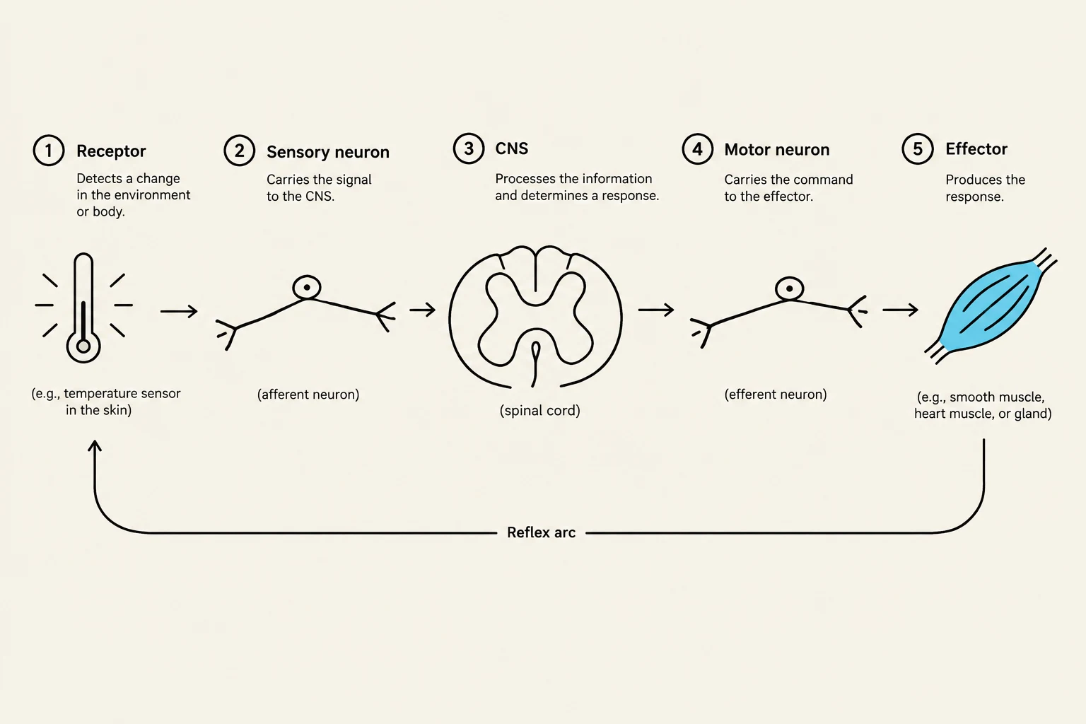
Through these complex reflex arcs—consisting of a receptor, sensory neuron, integrating center, motor neurons, and an effector—the autonomic nervous system ensures that functions like heart rate, blood pressure, and digestion are perfectly tuned to the body's immediate needs.
Diagram illustrating the components of a reflex arc, including receptor, sensory neuron, CNS, motor neuron, and effector.
Keep reading for free.
Create an account to finish this reading and make it part of your study routine.