T Cell and B Cell Receptor Complexes: Structure and Signaling
Explores the structure, function, and signaling pathways of T cell receptor (TCR) and B cell receptor (BCR) complexes, crucial for adaptive immunity. Covers antigen recognition, coreceptors, immune synapses, and cytokine receptor signaling via the JAK-STAT pathway.
The adaptive immune system relies on the ability of lymphocytes to recognize specific antigens with high precision. This recognition is mediated by specialized surface structures: the T cell receptor (TCR) complex and the B cell receptor (BCR) complex. While these receptors serve different roles—T cells generally respond to processed peptides presented by other cells, while B cells recognize intact antigens—they share fundamental principles of structure, clustering, and intracellular signal transduction. Understanding these complexes is essential for grasping how the immune system transitions from antigen recognition to a full-scale physiological response.
The T Cell Receptor (TCR) Complex
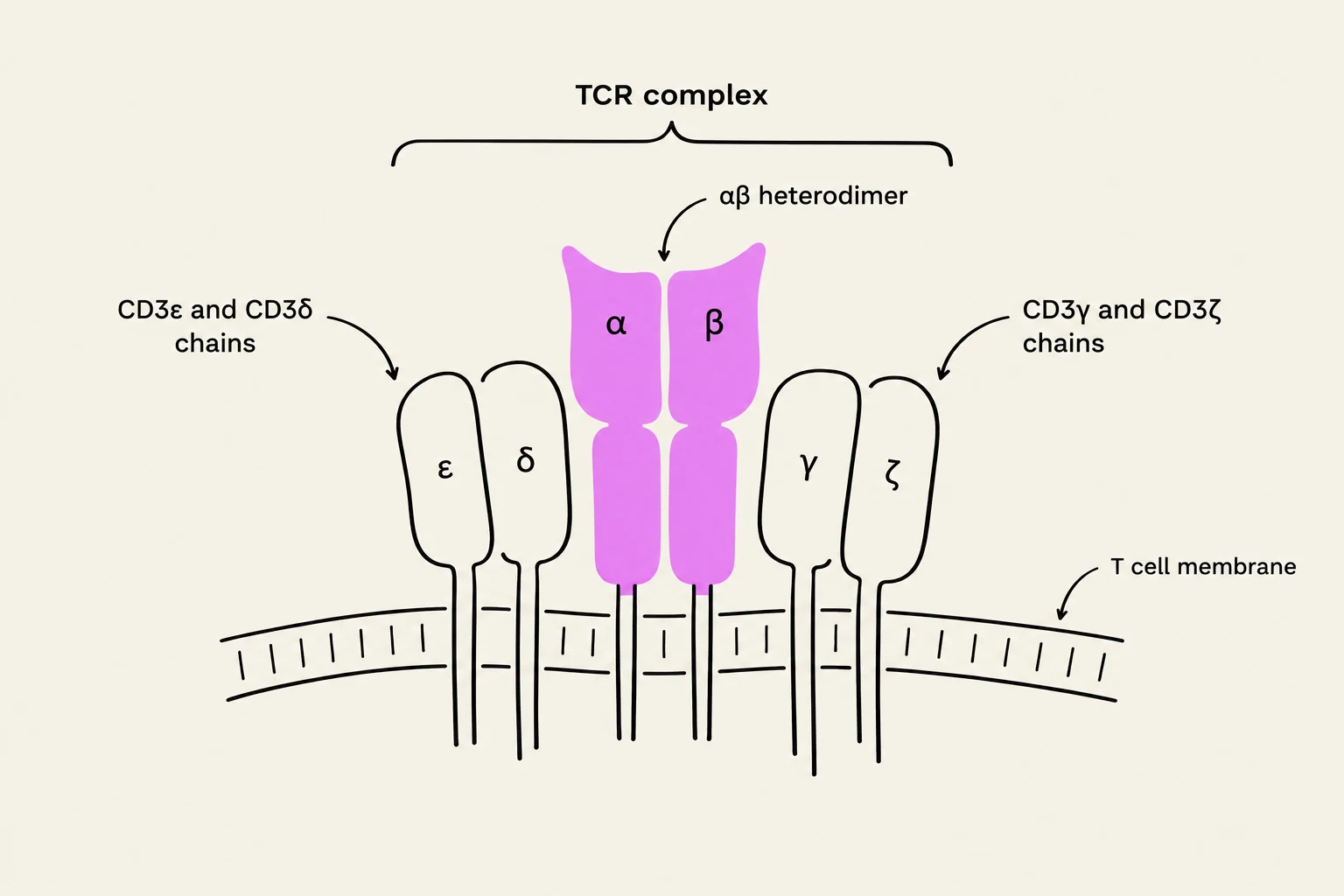
The TCR is not a solitary molecule but a multi-protein complex designed to convert extracellular binding events into intracellular signals. The core of this complex is the TCR itself, which is a heterodimer consisting of two transmembrane polypeptide chains covalently linked by disulfide bridges. In most T cells (αβ T cells), these chains are α and β, though γδ T cells utilize γ and δ chains. Each chain contains an immunoglobulin (Ig)-like N-terminal variable (V) domain containing hypervariable complementarity-determining regions (CDRs), a constant (C) domain, a hydrophobic transmembrane region, and a short cytoplasmic tail.
Because the cytoplasmic tails of the α and β chains are too short to transmit signals, the TCR must associate with auxiliary proteins: CD3 (composed of γ, δ, and ϵ chains) and the Protein ζ (zeta) chain. These associated proteins contain Immunoreceptor Tyrosine-based Activation Motifs (ITAMs), which are critical for the initiation of signaling.
Diagram of the T cell receptor (TCR) complex, illustrating its components including the αβ heterodimer and associated CD3 chains (CD3ε, CD3δ, CD3γ, and CD3ζ) embedded in the T cell membrane.
Antigen Recognition and Coreceptors
T cells do not recognize free-floating antigens. Instead, they recognize MHC-antigen-peptide complexes presented on the surface of Antigen-Presenting Cells (APCs). To ensure efficient signaling and specificity, T cells utilize coreceptors:
CD4: Found on helper T cells; it binds to Class II MHC molecules.
CD8: Found on cytotoxic T cells; it binds to Class I MHC molecules.
These coreceptors are members of the Ig superfamily and facilitate signaling by bringing the kinase LCK into close proximity with the TCR complex.
The Immune Synapse (SMAC)
When a T cell engages an APC, the contact area organizes into a highly structured region known as the Supramolecular Activation Cluster (SMAC), or the immune synapse. This "bull's eye" structure consists of three distinct concentric rings:
c-SMAC (central): Contains the TCR complex, CD4/CD8 coreceptors, and costimulatory receptors like CD28.
p-SMAC (peripheral): Formed by integrins (such as LFA-1/ICAM-1) that stabilize the physical bond between the cells.
d-SMAC (distal): The outermost region containing molecules like CD43, CD44, and CD45.
Exam Tip: Remember that the c-SMAC is the "signaling hub" where the actual TCR-MHC interaction occurs, while the p-SMAC provides the mechanical stability required to maintain that contact.
B Cell Receptor (BCR) Complex Signaling
While T cells look for "presented" peptides, B cells recognize antigens directly via the B cell receptor (BCR) complex. The BCR is essentially a membrane-bound antibody molecule, typically of the IgM or IgG isotype.
Similar to the TCR, the antibody portion of the BCR cannot signal on its own. It must be coupled with two signaling chains: Igα (CD79a) and Igβ (CD79b). When an antigen binds to the BCR, it triggers a cascade involving tyrosine kinases, most notably Spleen Tyrosine Kinase (SYK). This leads to several downstream pathways, including:
The RAS-MAP kinase pathway.
Phosphatidylinositol-specific phospholipase C (PLC) activation.
Protein kinase C β (PKC β) signaling.
B cell activation can be further enhanced by the B cell coreceptor complex (comprising CD21, CD19, CD81, and CD225) and complement proteins like C3d, which bridge innate and adaptive immunity.
Cytokine Receptors and the JAK-STAT Pathway
Cytokines are small, non-structural proteins (5–25 kDa) that act as chemical messengers between immune cells. They are categorized into families including interleukins, interferons, tumor necrosis factors, and chemokines. Cytokines exhibit unique functional properties:
Pleiotropy: One cytokine affects many different cell types.
Redundancy: Different cytokines produce the same effect.
Synergy: Two cytokines working together produce a greater effect than the sum of their parts.
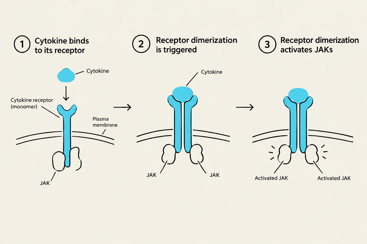
This diagram illustrates the process of cytokine signaling, where a cytokine molecule binds to its receptor on the plasma membrane, triggering receptor dimerization and subsequent activation of JAK (Janus kinase) proteins.
Signaling Mechanisms
All cytokine receptors share a common architecture: an extracellular binding domain, a transmembrane domain, and a cytoplasmic domain for signal initiation.
A primary mechanism for cytokine signaling is the JAK-STAT pathway:
Binding: The cytokine binds to the receptor, causing the receptors to dimerize.
Activation: This dimerization activates Janus kinases (JAKs) associated with the receptor tails.
Phosphorylation: JAKs phosphorylate the receptor, creating docking sites for STAT (Signal Transducers and Activators of Transcription) proteins.
Transcription: STAT proteins are phosphorylated, form dimers, and move into the nucleus to activate specific genes.
Clinical Relevance
Because the JAK-STAT pathway is so central to inflammation, it is a major target for drug therapy. JAK inhibitors (such as tofacitinib and baricitinib) are used to treat autoimmune conditions like rheumatoid arthritis and psoriasis. However, clinicians must monitor for side effects like increased infection risk and cytopenias due to the systemic nature of these pathways.
Summary Table: TCR vs. BCR Comparison
Feature
T Cell Receptor (TCR)
B Cell Receptor (BCR)
Antigen Type
Processed peptides on MHC
Intact antigens
Core Structure
αβ or γδ heterodimer
Antibody (IgM/IgG)
Signaling Chains
CD3 and Protein ζ
Igα and Igβ
Key Kinases
LCK
SYK
Keep reading for free.
Create an account to finish this reading and make it part of your study routine.