


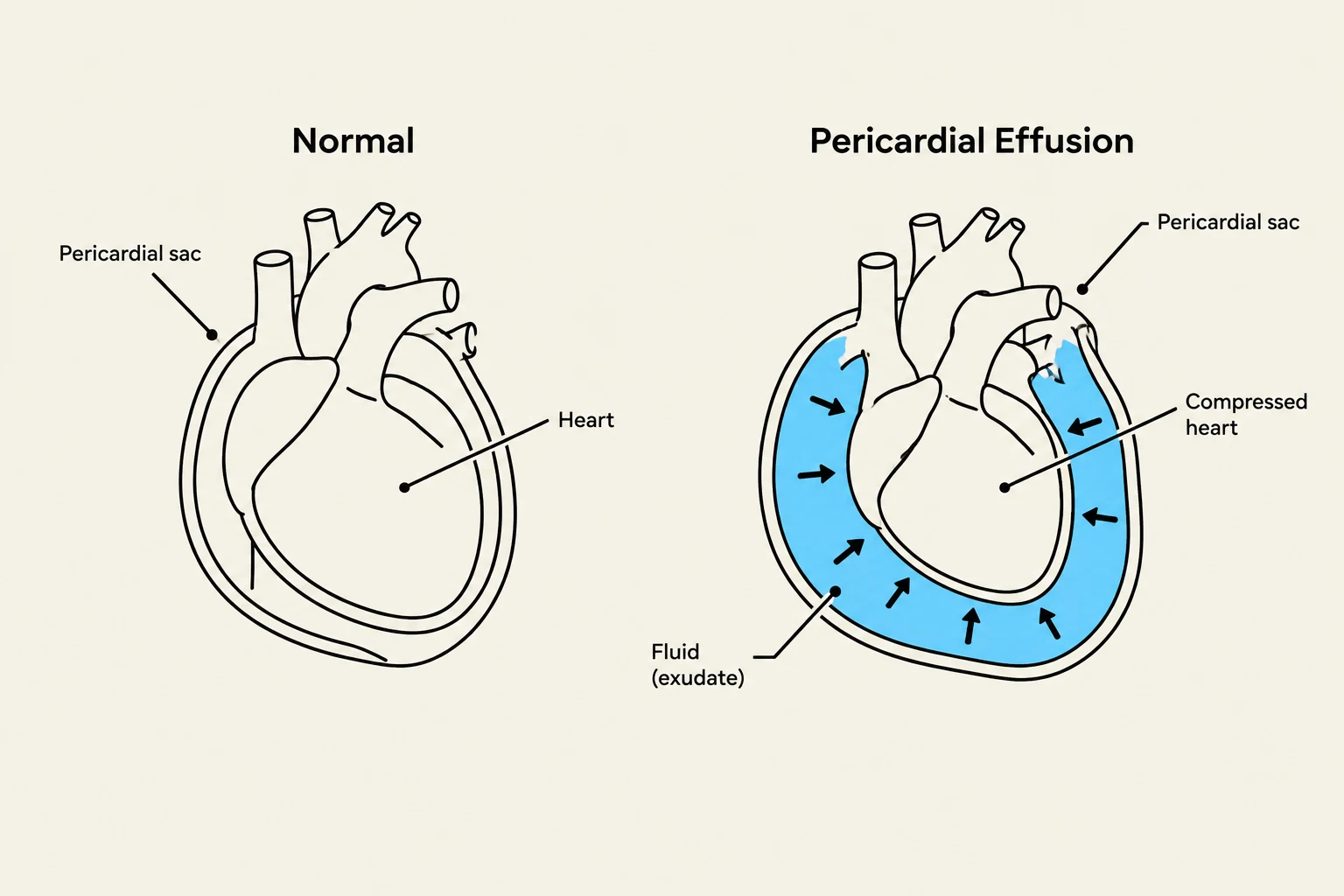
The heart is protected by a specialized double-layered membrane known as the pericardium. This structure consists of an outer parietal layer (which is fibrous) and an inner visceral layer that is attached directly to the heart. Between these two layers lies the pericardial space, which normally contains a small amount of pericardial fluid to facilitate smooth movement during the cardiac cycle.
Pericarditis occurs when this membrane becomes inflamed due to infection, trauma, or underlying conditions like renal failure or autoimmune diseases. This inflammation can lead to the accumulation of an exudate (excess fluid) or the formation of a thick, fibrous sac that restricts the heart's movement. When this fluid accumulates excessively, it can lead to a life-threatening condition called cardiac tamponade, where the heart is compressed from the outside.
Primary Clinical Symptoms
Patients suffering from pericarditis often present with distinct physical sensations that reflect the underlying inflammation and mechanical restriction of the heart.
Retrosternal Pain
One of the most hallmark symptoms is severe retrosternal pain. This pain is localized specifically behind the sternum (the breastbone). Because the inflammation involves nerve irritation in the pericardial layers, the pain can be sharp and may change in intensity based on the patient's position or breathing patterns.
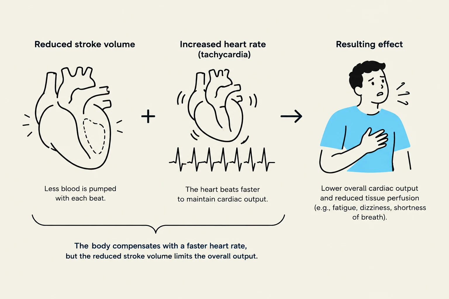
Tachycardia and Dyspnea
As the inflammation progresses, it often impairs the heart's ability to contract and relax properly. This leads to a significant reduction in cardiac output—the total volume of blood pumped by the left ventricle per minute.
To understand how the body reacts, we must look at the relationship between stroke volume and heart rate. Cardiac output () is calculated as follows:
Keep reading for free.
Create an account to finish this reading and make it part of your study routine.
Continue reading freeAlready have an account? Log in
Updated