


Surgical staging is a cornerstone in the management of endometrial cancer, serving as the primary method to determine the extent of disease spread and guide subsequent treatment decisions. The ultimate goal of staging is to accurately identify lymph node metastasis, which is a critical prognostic factor that dictates whether a patient requires adjuvant therapy, such as radiation or systemic chemotherapy.
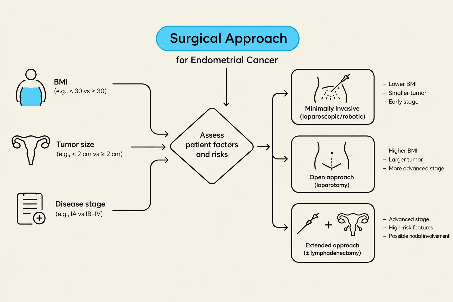
In recent years, the paradigm has shifted from traditional, more invasive lymphadenectomy toward more precise, targeted techniques and minimally invasive surgical approaches. This evolution aims to balance oncologic accuracy—ensuring no cancer is missed—with the reduction of surgical morbidity and complications.
Sentinel Lymph Node (SLN) Mapping and Ultrastaging
For many patients, particularly those with low-to-intermediate risk disease that appears confined to the uterus, identifying the "sentinel" node—the first node(s) to receive lymphatic drainage from the tumor—can replace the need for a full lymphadenectomy.
SLN Mapping involves using specialized dyes or radioactive tracers to trace the primary lymphatic pathway. When performed according to established surgical algorithms, this technique demonstrates high sensitivity (up to 97.2%) and a very low false-negative rate (less than 5%). While its use was once debated for high-risk histologies, current evidence suggests that SLN mapping provides acceptable accuracy even in aggressive cases such as serous carcinoma, clear cell carcinoma, or carcinosarcoma.
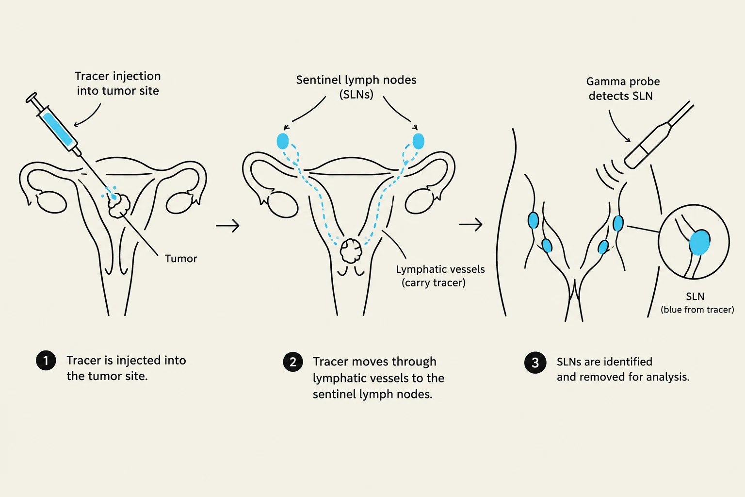
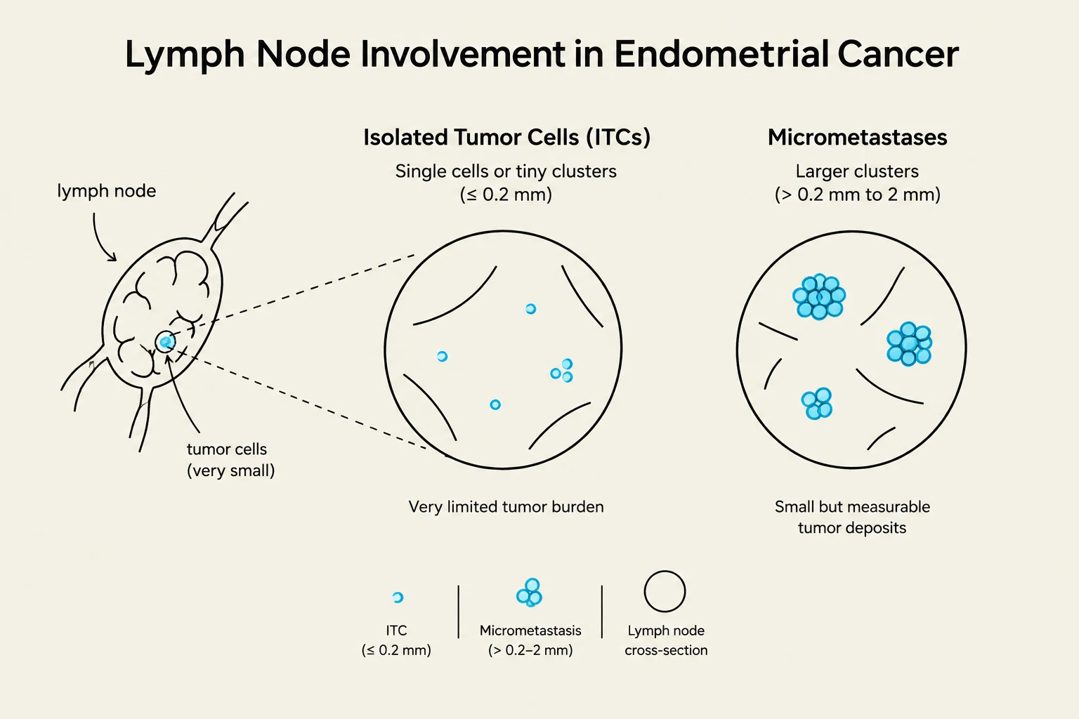
To ensure that even the smallest clusters of cancer cells are not overlooked, pathologists employ SLN ultrastaging. This process involves serial sectioning of the identified nodes and the use of immunohistochemistry (IHC) to detect microscopic disease. While ultrastaging is highly effective, it can lead to "upstaging" (finding cancer that was not visible on standard inspection) in approximately 5% to 15% of patients.
Keep reading for free.
Create an account to finish this reading and make it part of your study routine.
Continue reading freeAlready have an account? Log in
Updated