


The management of cervical cancer has evolved from standardized surgical resection to a highly personalized approach driven by surgical precision and molecular profiling. For early-stage disease, the goal is curative resection with minimal morbidity, whereas advanced or recurrent disease increasingly relies on targeted systemic therapies guided by specific biomarkers. Understanding the interplay between surgical staging, pathologic risk stratification, and genomic testing is essential for modern oncologic practice.
Surgical Management of Early-Stage Disease
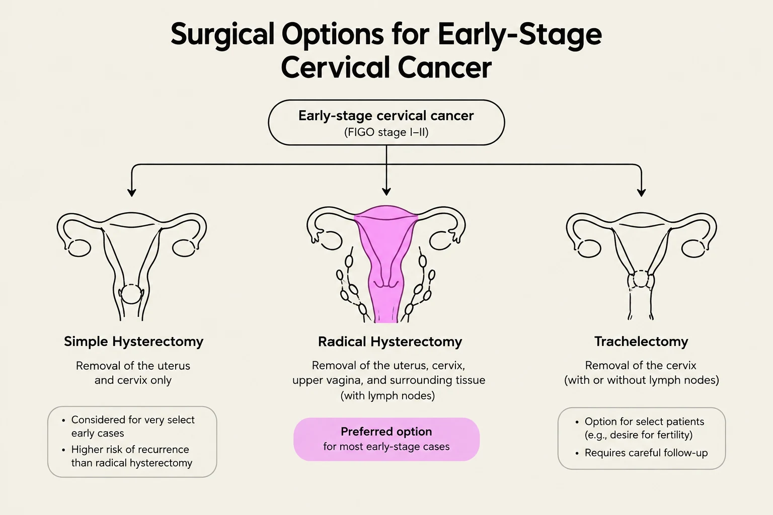
For patients with early-stage cervical cancer (specifically FIGO stages IB1, IB2, IB3, IIA1, and IIA2), the primary surgical objective is achieving wide resection margins to prevent local recurrence.
Radical Hysterectomy (Type C) is the preferred approach for these stages due to its ability to provide wider margins compared to simpler hysterectomies. However, clinical decision-making must account for the specific FIGO stage; for instance, in the United States, definitive chemoradiation is often the preferred route for more extensive lesions, such as FIGO IB3 and stage IIA2. In cases where disease has recurred but remains confined to the central pelvis, pelvic exenteration may be considered as a potentially curative surgical option.
Clinical Pearl: While Minimally Invasive Surgery (MIS) is often favored for faster recovery, the LACC trial demonstrated that MIS is associated with lower disease-free survival (DFS) and overall survival (OS) compared to open abdominal radical hysterectomy.
Lymph Node Assessment and Staging
Accurate nodal staging is critical for determining the necessity of adjuvant therapy.
Sentinel Lymph Node (SLN) Mapping
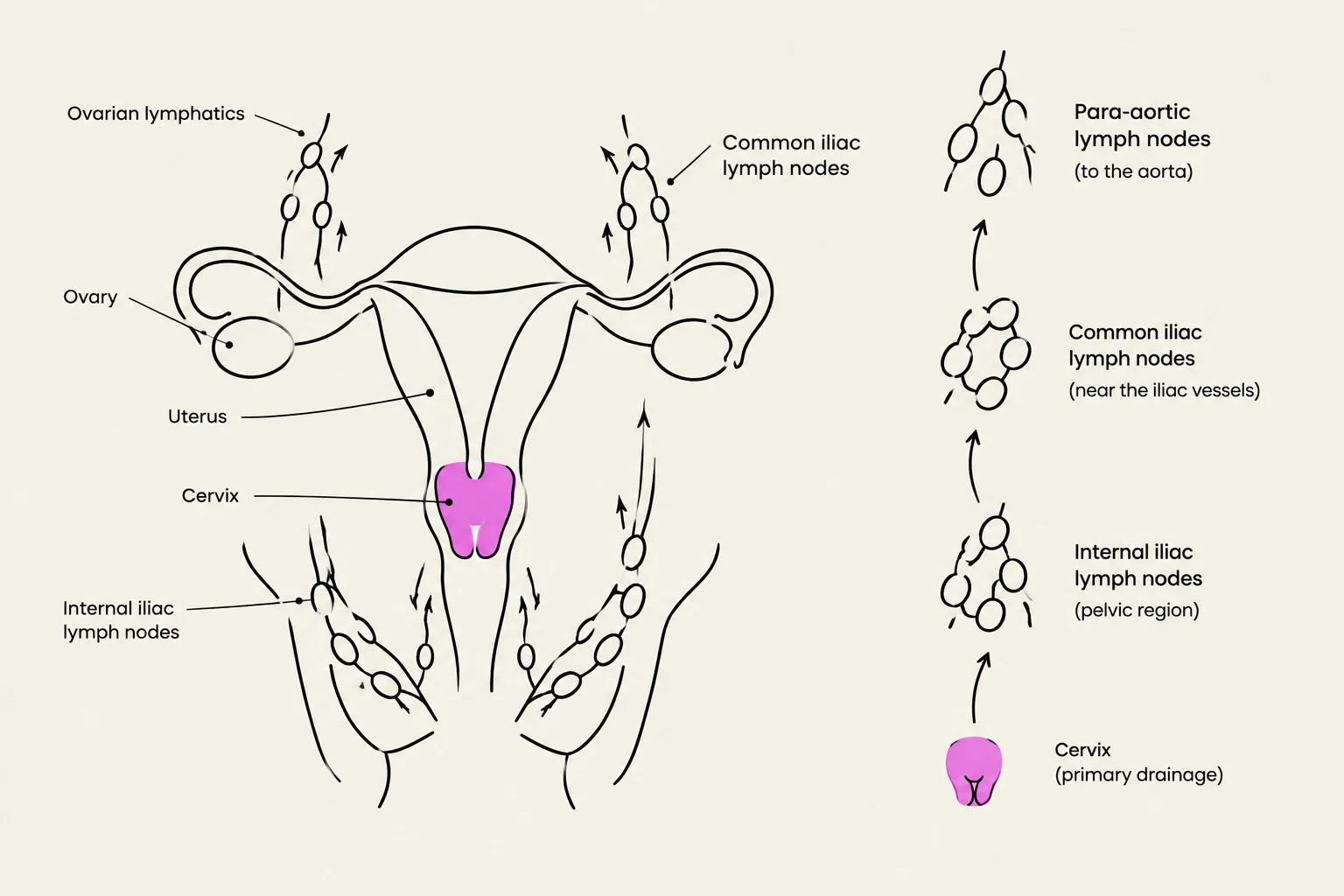
To reduce the morbidity associated with extensive pelvic lymphadenectomy, Sentinel Lymph Node (SLN) mapping is utilized in early-stage cases. Using tracers such as Indocyanine green (ICG), clinicians can identify the first nodes to receive drainage from the tumor. Detection rates for SLN are high, approximately 89%–92%. It is important to note that ultrastaging is a critical component of this process to detect micrometastases and isolated tumor cells that might be missed by standard microscopy.
Keep reading for free.
Create an account to finish this reading and make it part of your study routine.
Continue reading freeAlready have an account? Log in
Updated