Superficial Skeletal Muscles of the Human Body: Anatomy and Function
An overview of the superficial skeletal muscles of the human body, detailing their locations and primary functions in the head, neck, trunk, and limbs. Covers muscles involved in facial expression, mastication, swallowing, and locomotion.
The human body is equipped with a complex system of skeletal muscles that allow for everything from subtle facial expressions to powerful locomotive movements. When studying anatomy, it is helpful to categorize these muscles by their location and their primary functional roles. The "superficial" muscles are those located closest to the surface of the body, making them primary movers and key players in visible physical actions. This article explores the superficial muscle groups of the head, neck, trunk, and limbs.
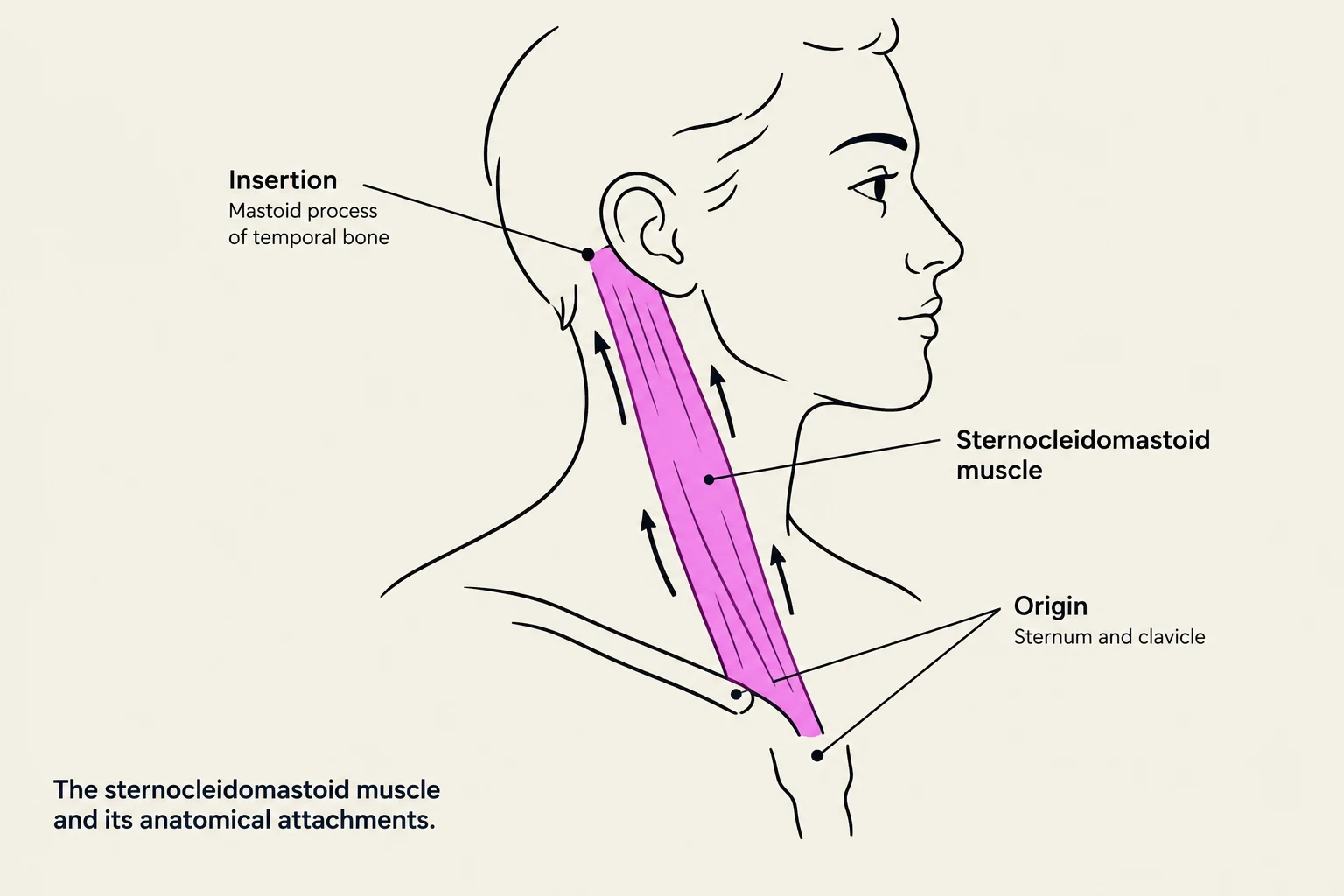
An illustration showing the sternocleidomastoid muscle, its origin on the sternum and clavicle, and its insertion on the mastoid process of the temporal bone.
Head and Neck Muscles
The muscles of the head and neck are specialized for communication, nutrition, and airway management.
Facial Expression and Neck
Facial expressions are driven by superficial muscles that manipulate the skin and mouth. The orbicularis oris is essential for mouth closure and shaping the oral cavity. Working alongside it is the buccinator, the principal muscle of the cheek, which helps keep food positioned between the teeth during chewing.
In the neck, the platysma and the sternocleidomastoid are prominent superficial muscles. The sternocleidomastoid is named for its attachments: the sternum and clavicle (origins) and the mastoid process (insertion).
Mastication (Chewing)
Chewing is made possible by the mastication muscles. The temporalis helps elevate and retract the jaw, while the masseter is a powerful muscle used to elevate the jaw. While deeper muscles like the medial and lateral pterygoids assist, these superficial muscles are the primary drivers of jaw movement.
Hyoid Movement: Swallowing and Speaking
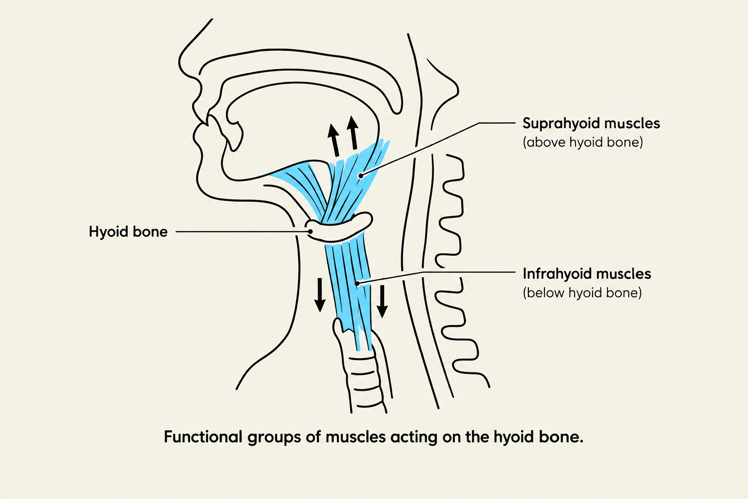
Diagram illustrating the suprahyoid and infrahyoid muscles acting on the hyoid bone.
The hyoid bone serves as an anchor for muscles that facilitate swallowing, speaking, and breathing. These are divided into two functional groups:
Suprahyoid muscles: These include the mylohyoid, stylohyoid, and digastric. They work to elevate the hyoid bone and the floor of the mouth, helping to push food toward the pharynx.
Infrahyoid muscles: These include the omohyoid and sternohyoid. Their primary role is to depress the hyoid and the larynx.
Study Tip: Remember that "supra-" means above and "infra-" means below. Suprahyoid muscles pull the hyoid upward, while infrahyoid muscles pull it downward.
Upper Body and Limbs
The muscles of the upper body are organized to provide both stability to the torso and a wide range of motion to the arms.
Shoulder and Trunk
The shoulder girdle is moved by several key superficial muscles. The trapezius is responsible for elevating, retracting, or depressing the scapula. The deltoid is a multi-functional muscle that can flex, rotate, adduct, abduct, and extend the shoulder. For movements involving the arm itself, the pectoralis major assists in adduction, rotation, flexion, and extension, while the latissimus dorsi aids in extending, adducting, and rotating the shoulder.
The trunk provides the core stability necessary for posture and movement. The serratus anterior helps with the upward rotation and stabilization of the scapula. The abdominal wall is composed of the rectus abdominis (responsible for trunk flexion and pelvic tilt) and the external obliques (which assist in trunk flexion, rotation, and abdominal compression).
Arm and Forearm
The muscles of the arm are categorized by their position:
Anterior Arm: This group is dominated by the biceps brachii, which facilitates elbow flexion, shoulder flexion, and forearm supination. The brachialis also assists in elbow flexion, while the brachioradialis supports forearm rotation and elbow stabilization.
Posterior Arm: The triceps brachii is the primary muscle here, consisting of three heads (lateral, long, and medial) that work together to extend the elbow and shoulder.
The forearm muscles manage the movement of the wrist and fingers:
Anterior Forearm (Flexors): These muscles, such as the pronator teres, flexor carpi radialis, palmaris longus, and flexor carpi ulnaris, are responsible for forearm pronation and wrist flexion.
Posterior Forearm (Extensors): These muscles, including the extensor carpi radialis group and the extensor digitorum, manage the extension of the wrist and the straightening of the fingers.
Hand Muscles
While the specific names of every small muscle are often detailed in advanced study, the superficial muscles of the hands generally serve to protect the digits, enhance grip strength, stabilize the fingers, and generate torque.
Lower Body and Legs
The lower limb muscles are designed for weight-bearing, balance, and locomotion.
Anterior Lower Leg
The muscles on the front of the lower leg primarily manage the position of the foot. The tibialis anterior is the primary muscle for dorsiflexion (lifting the foot) and inversion. Other muscles like the extensor digitorum longus and extensor hallucis longus assist in extending the toes and aiding in ankle dorsiflexion.
Posterior Lower Leg (The Calf)
The posterior compartment is largely responsible for plantarflexion (pointing the toes downward). This area is often referred to as the "calf" and includes the gastrocnemius (which has medial and lateral heads) and the soleus. The plantaris also contributes to plantarflexion and knee flexion.
Thigh and Hip
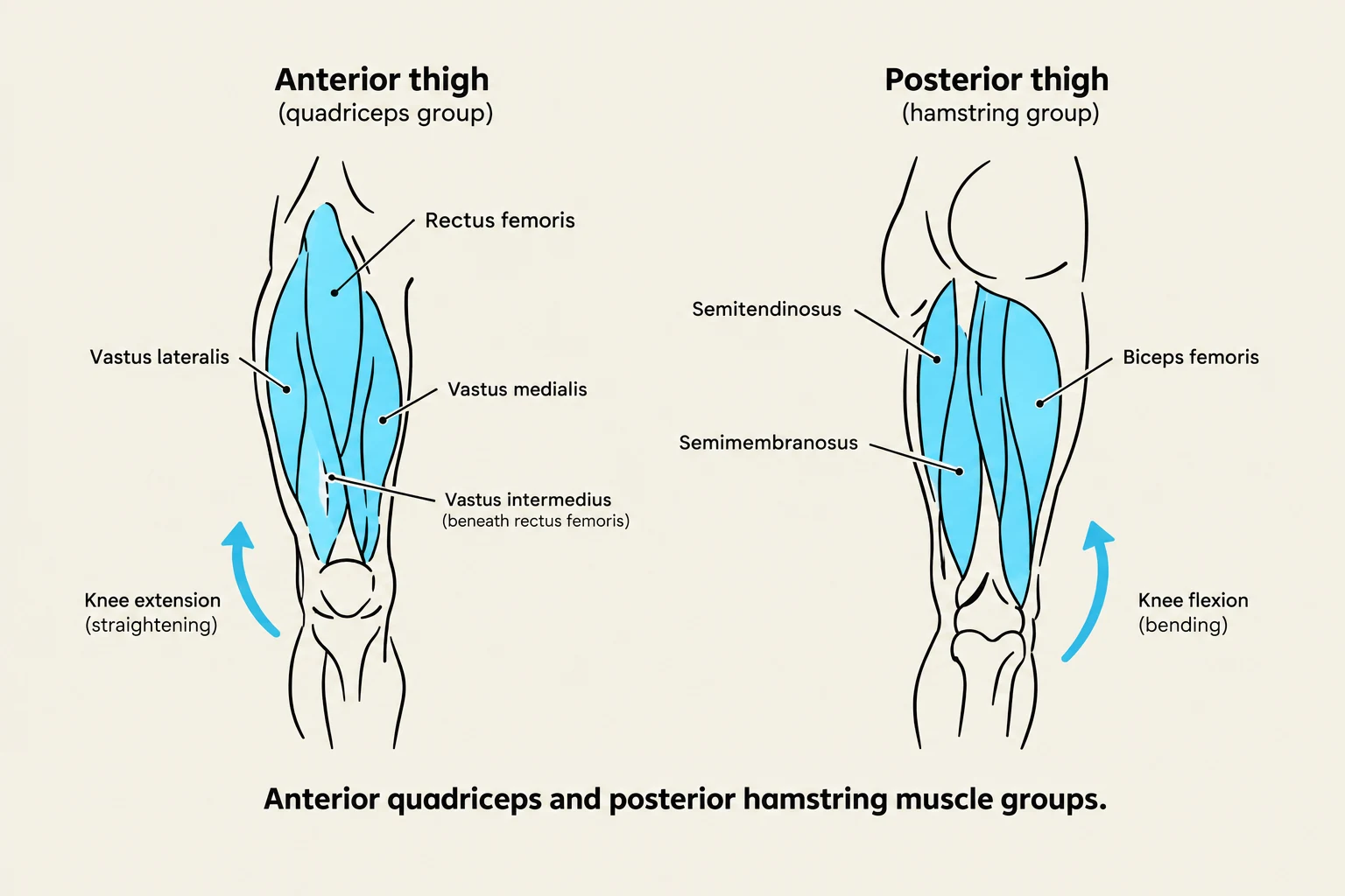
Anterior quadriceps and posterior hamstring muscle groups.
The thigh contains large muscle groups that drive movement:
Anterior Thigh: The quadriceps group (comprising the rectus femoris, vastus lateralis, vastus medialis, and vastus intermedius) is essential for knee extension.
Posterior Thigh: The hamstrings (semitendinosus, semimembranosus, and biceps femoris) are responsible for knee flexion and hip extension.
Hip Stabilizers: Muscles like the sartorius, adductor longus, and tensor fasciae latae contribute to hip flexion, abduction, and rotation, while also providing stability to the knee and pelvis.
Exam Tip: When identifying muscles, look at the name. For example, the sternocleidomastoid is named after its origin (sternum/clavicle) and its insertion (mastoid process).
Keep reading for free.
Create an account to finish this reading and make it part of your study routine.