


The integumentary system serves as the body's first line of defense, providing a protective barrier against environmental hazards, regulating temperature, and facilitating sensory perception. Understanding the complex architecture of the skin—from its superficial layers to its deep connective tissues—is essential for identifying various dermatological conditions, ranging from common inflammatory responses to serious tissue injuries.
Layers of the Skin
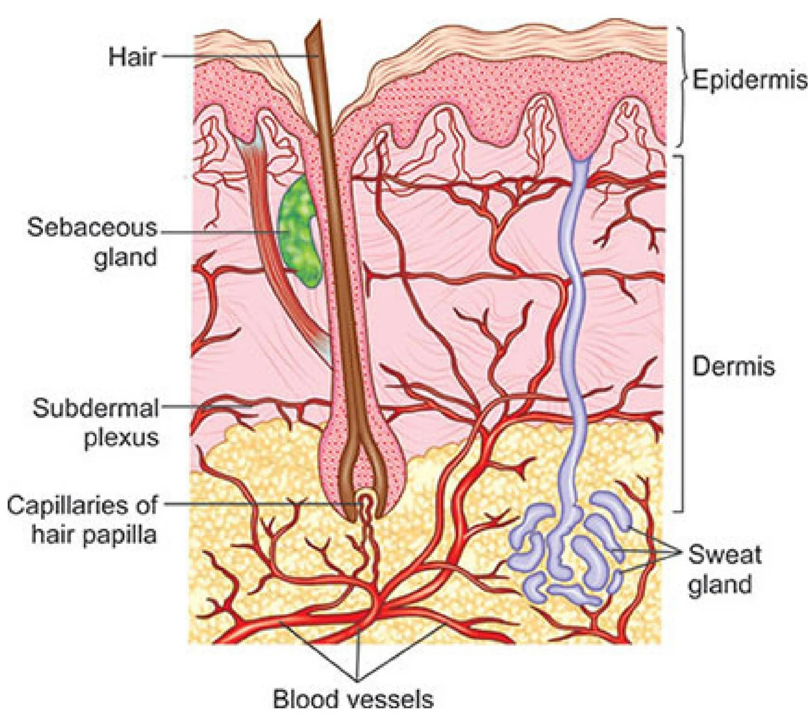
The skin is organized into three primary layers, each with distinct histological characteristics and functions.
Epidermis
The epidermis is the superficial outer layer of the skin. It acts as a waterproof barrier and provides our primary protection against pathogens and UV radiation.
Dermis
Located beneath the epidermis, the dermis is often referred to as the "true skin". It contains much of the skin's structural integrity, including connective tissue, blood vessels, and nerves. While blood supply to the skin is limited to the papillary capillaries, the sympathetic nervous system plays a critical role in regulating skin temperature through vasoconstriction and vasodilation.
Hypodermis (Subcutaneous Layer)
The hypodermis is the lowest layer of connective tissue. It serves as a reservoir for energy and provides cushioning for underlying structures. This layer is composed of:
- Macrophages and fibroblasts
- Fat cells (adipocytes)
- Fine muscles and blood vessels
- Lymphatics and hair follicle roots

Dermal Appendages
Dermal appendages are specialized structures that extend from the skin to perform specific functions such as thermoregulation, sensation, and protection.
- Nails and Hair: Provide protection and sensory input.
- Sebaceous Glands: Produce oil (sebum) to lubricate the skin and hair.
- Sweat Glands:
- Eccrine sweat glands: These are prominent in the hands, soles of the feet, and the forehead. They are essential for thermoregulation.
- Apocrine sweat glands: Located near hair follicles in the axillae (armpits), scalp, face, abdomen, and genitalia.

Keep reading for free.
Create an account to finish this reading and make it part of your study routine.
Continue reading freeAlready have an account? Log in
Updated