Explores the hierarchical organization of skeletal muscle, from the myofiber to the sarcomere, detailing the molecular interactions that generate force through actin and myosin filaments. Covers excitation-contraction coupling and factors influencing muscle force generation.
To understand how a human being lifts a heavy weight or maintains posture, we must zoom in past the visible muscle and into the microscopic world of proteins. At the heart of every movement is a highly organized, repeating structure called the sarcomere. This article explores the hierarchical organization of skeletal muscle, from the entire muscle fiber down to the molecular interactions that generate force.
The hierarchical organization of skeletal muscle from macro to micro, illustrating the progression from a whole muscle to myofibrils and sarcomeres, detailing the actin and myosin filaments within a sarcomere.
The Hierarchy of Muscle: From Myofiber to Sarcomere
Skeletal muscle is organized into a complex hierarchy of structures. At the largest cellular level, we have the myofiber (or muscle cell). Unlike most cells in the body, myofibers are multinucleated, meaning they contain many nuclei located on the periphery of the cell. This high number of nuclei is necessary to support the massive amount of gene transcription and protein synthesis required for such a large, highly metabolic cell.
Within a single myofiber, there are numerous rod-like organelles called myofibrils that run the length of the cell. These myofibrils are the actual contractile elements of the muscle. If you were to look at a myofibril under an electron microscope, you would see it is composed of even smaller protein filaments called myofilaments.
The functional unit of the myofibril—and indeed the entire muscle—is the sarcomere. Sarcomeres are arranged in series, end-to-end, within the myofibril. The repeating pattern of these sarcomeres gives skeletal muscle its characteristic "striated" or striped appearance under a microscope.
Microstructure of the Myofiber
Before we look at the sarcomere itself, we must understand the environment that supports it. The myofiber is not just a bag of proteins; it is a sophisticated machine with specialized compartments:
Sarcolemma: The cell membrane that surrounds the myofiber.
Sarcoplasm: The cytoplasm of the cell, which is packed with mitochondria for ATP production and ribosomes for protein translation.
Sarcoplasmic Reticulum (SR): A specialized network that acts as a storage warehouse for calcium ions (Ca2+). The release and reuptake of calcium are what turn muscle contraction "on" and "off".
T-tubules: Extensions of the sarcolemma that tunnel deep into the cell, carrying electrical signals (action potentials) from the surface directly to the SR.
Extracellular Matrix (ECM): A network of connective tissue, primarily collagen, that provides structural support and helps transmit the force generated by the sarcomeres to the rest of the body.
The Repair Kit: Satellite Cells
Every myofiber has its own "repair kit" known as the satellite cell niche. Located between the sarcolemma and the basal lamina, satellite cells are muscle stem cells that reside in a dormant, or "quiescent," state. When a muscle is injured or undergoes the stress of resistance training, these cells are activated, proliferate, and eventually fuse with the myofiber to donate their nuclei, helping the muscle repair or grow (hypertrophy).
The Anatomy of a Sarcomere
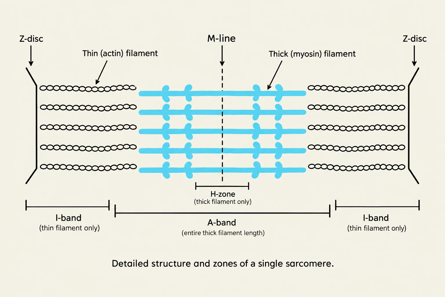
Detailed structure and zones of a single sarcomere.
The sarcomere is composed of two primary types of myofilaments: thick filaments and thin filaments.
The Filaments
Thick Filaments: Comprised primarily of the protein myosin. Myosin molecules have long tails and protruding "heads" that act as molecular motors to pull on the thin filaments.
Thin Filaments: Comprised primarily of the protein actin. Actin filaments are spherical proteins joined in a helical shape that provide binding sites for myosin.
To regulate contraction, two additional proteins are attached to the actin:
Tropomyosin: A filament protein that blocks the myosin-binding sites on actin when the muscle is at rest.
Troponin: A globular protein bound to tropomyosin. It acts as the "lock" that responds to calcium.
Structural Anchors and Zones
A single sarcomere is defined by specific boundaries and landmarks:
Z-discs: The boundaries of the sarcomere. These anchor the thin filaments at each end.
M-line: The very center of the sarcomere, which serves as the anchor point for the thick filaments.
Titin: A massive, spring-like protein that connects the thick filaments to the Z-discs, providing structural stability and elastic recoil.
When viewing a sarcomere, we categorize regions based on the arrangement of these filaments:
A-band: The entire length of the thick filament, including the area where thick and thin filaments overlap.
I-band: The region containing only thin filaments; it spans across the Z-disc into the adjacent sarcomere.
H-zone: The central region of the A-band that contains only thick filaments (no overlap with actin).
Excitation-Contraction Coupling
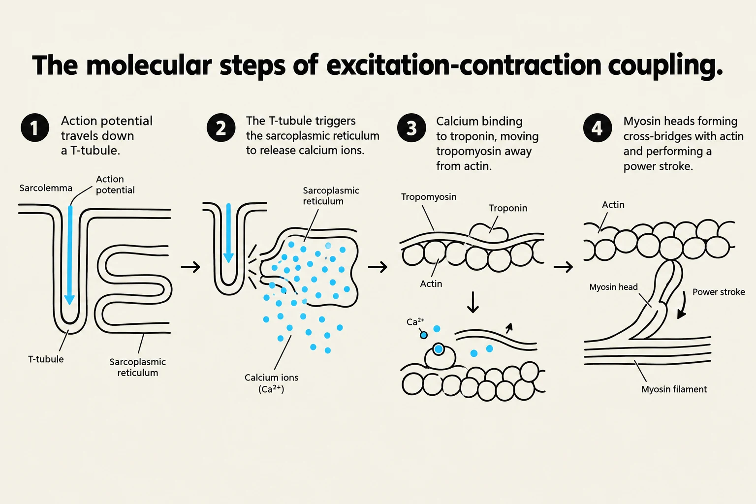
Diagram illustrating the four molecular steps of excitation-contraction coupling in muscle tissue, involving action potentials, calcium ion release, and the interaction of actin and myosin filaments.
How does a thought in your brain become a physical movement? This process is called excitation-contraction coupling.
Excitation: A signal from a motor neuron reaches the neuromuscular junction (NMJ), the interface between the neuron and the muscle fiber. This triggers an electrical impulse (action potential) that travels across the sarcolemma and down into the T-tubules.
Calcium Release: The electrical signal in the T-tubules triggers the sarcoplasmic reticulum (SR) to release a flood of calcium ions (Ca2+) into the sarcoplasm.
Cross-Bridge Formation: The Ca2+ binds to troponin, causing a conformational change that moves tropomyosin away from the binding sites on the actin filament.
Contraction: With the sites exposed, the myosin heads bind to the actin, forming cross-bridges. Using energy from ATP, the myosin heads perform a "power stroke," pulling the actin filaments toward the M-line.
Exam Tip: What happens during contraction?
As the muscle contracts and the sarcomere shortens:
The Z-discs move closer together.
The I-band shortens (gets smaller).
The H-zone shortens or disappears entirely.
The A-band length remains constant (because the length of the myosin filament does not change).
Factors Influencing Force Generation
The amount of force a muscle produces is not constant; it depends on several physiological and mechanical factors:
Length of the muscle fiber: The degree of overlap between thick and thin filaments determines how many cross-bridges can form. There is an "optimal length" (L0) where the muscle produces maximal force.
Frequency of stimulation: A single stimulus produces a twitch. If stimuli arrive rapidly, the muscle undergoes summation. If the frequency is high enough that the muscle cannot relax between stimuli, it reaches fused tetanus, the state of maximal sustained force.
Muscle fiber recruitment: The nervous system can increase force by activating more motor units (more muscle fibers).
Muscle Environment: Factors like temperature, pH (acidity), hydration, and nutrient availability all impact the efficiency of the contractile machinery.
Keep reading for free.
Create an account to finish this reading and make it part of your study routine.