An overview of the respiratory system's structure and function, including conductive and respiratory airways, defense mechanisms, gas exchange across the alveolar-capillary membrane, and the mechanics of the pleura.
The respiratory system is a complex network designed to facilitate the vital exchange of gases between the atmosphere and the blood. Beyond the primary task of oxygenating blood and removing carbon dioxide, the lungs serve as a critical interface for environmental defense, blood filtration, and metabolic regulation. To achieve these diverse functions, the system is organized into two distinct functional zones: the conductive airways and the respiratory airways.
The Conductive Airways and Anatomical Dead Space
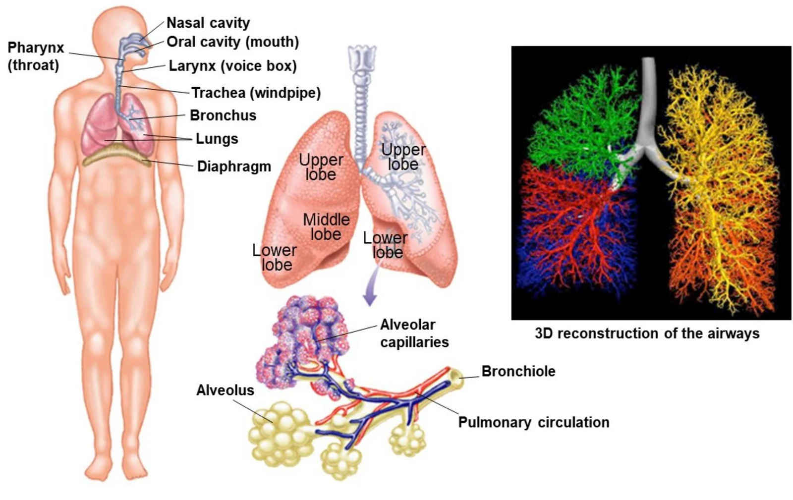
The respiratory tree begins with the upper respiratory tract, which prepares inhaled air by warming it to body temperature (37°C), humidifying it, and filtering out large particles via the nose and sinuses.
The first 16 generations of the airway are classified as conductive airways. These structures serve as conduits that transport air into and out of the deeper lung tissues. Because no gas exchange occurs within these generations, this region is referred to as the anatomical dead space.
Diagram illustrating the human respiratory system, including the lungs, airways, alveoli, and pulmonary circulation.
Structural Support and Regulation
To prevent the airways from collapsing during the pressure changes of breathing, the trachea and major bronchi are reinforced with hyaline cartilage. This cartilage is composed of a Type II collagen proteoglycan matrix that lacks direct blood vessels and nerves. Within this matrix, specialized cells called chondrocytes are responsible for secreting the extracellular matrix, ensuring the airway maintains both strength and flexibility.
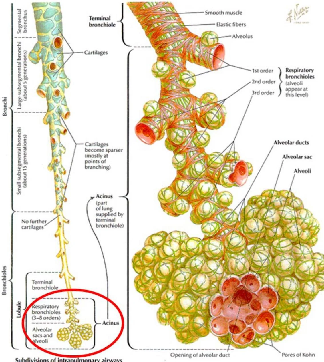
As the airways branch deeper into the lungs, the amount of cartilage decreases, and the proportion of smooth muscle increases. This allows for the regulation of airway diameter through autonomic nervous system control:
Parasympathetic stimulation (via acetylcholine) causes the smooth muscles to constrict.
Sympathetic stimulation (via β2 receptors and norepinephrine) causes the smooth muscles to dilate.
Exam Tip: Remember the inverse relationship between cartilage and smooth muscle. As you move from the trachea toward the bronchioles, cartilage decreases while smooth muscle increases.
Defense Mechanisms: The Mucociliary Escalator
Because the lungs are the primary interface between the body and the external environment, they require robust defense mechanisms to remove pathogens and dust.
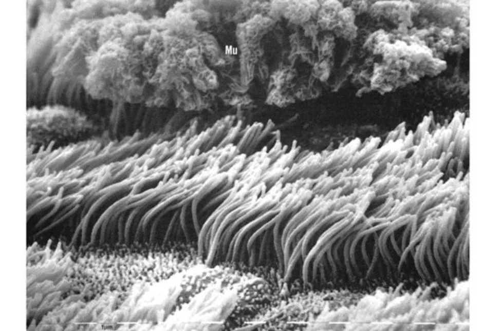
In the conducting zone, goblet cells produce mucus (glycoproteins/mucins) that traps large particles. The cells lining the respiratory tree down to the terminal bronchioles possess cilia that beat synchronously. This coordinated movement creates the mucociliary escalator, a process that carries the mucus film upward toward the trachea to be cleared.
Scanning electron micrograph of cilia and mucus on the surface of the respiratory epithelium.
When particles are too small to be trapped by mucus or reach the deepest parts of the lung, the body relies on cellular defense:
Large particles (>5 μm): Usually trapped in the nasopharynx via impaction.
Medium particles (1-5 μm): Settle in small airways via sedimentation.
Small particles (<0.1 μm): Reach the alveoli and are engulfed by alveolar macrophages.
The Respiratory Zone and Gas Exchange
The final 7 generations of the airways (generations 17–23) constitute the respiratory airways. In this zone, the total cross-sectional area increases exponentially, causing the velocity of airflow to drop significantly. This low velocity allows diffusion to become the primary mechanism for moving gases.
The Alveolar-Capillary Membrane
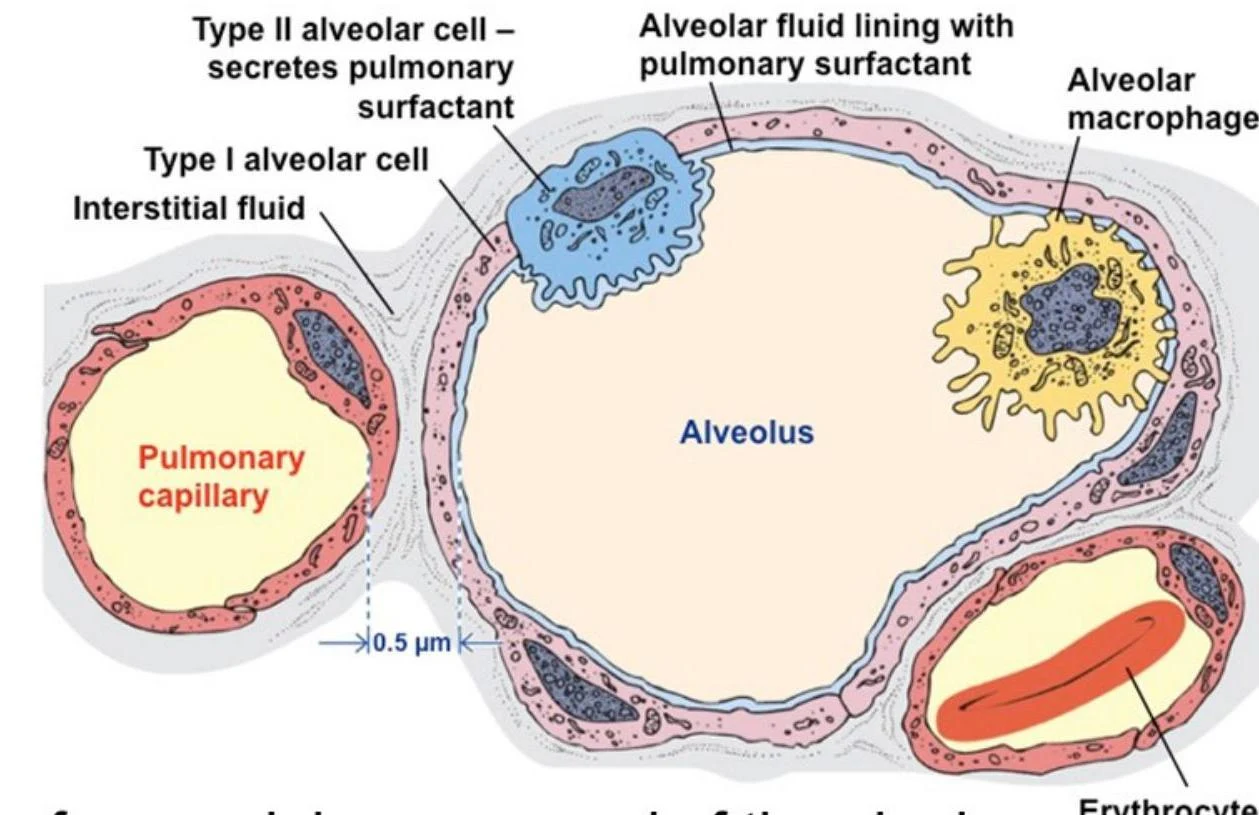
The core function of the respiratory zone is gas exchange. This occurs across the extremely thin alveolar-capillary membrane, which measures only approximately 0.2 to 0.5 μm in thickness.
Diagram illustrating the subdivisions of intrapulmonary airways, including bronchioles, alveolar ducts, alveolar sacs, and alveoli, with labels for associated structures like smooth muscle, elastic fibers, and cartilage.
The alveolar surface is composed of a highly specialized cellular landscape:
Type I epithelial cells (Aerocytes): These are thin, squamous cells that cover roughly 90–95% of the alveolar surface, providing the minimal distance required for rapid gas diffusion.
Type II pneumocytes: These larger, cuboidal cells are interspersed among the Type I cells. Their primary role is to secrete surfactant, a substance that reduces surface tension and prevents alveolar collapse.
gCap cells: These are stem-like endothelial cells located in the capillaries that can divide to regenerate aerocytes.
Diagram illustrating the structure of an alveolus and surrounding pulmonary capillaries, showing alveolar cells, macrophages, and erythrocytes.
Clinical Note: Type II pneumocytes are not just for surfactant; they also act as progenitor cells. If the lung is injured, Type II cells can divide to regenerate both Type I and Type II epithelial cells, though this process can take several weeks.
Mechanics of the Pleura and Pneumothorax
The lungs are contained within the pleural membranes, which consist of the visceral pleura (adjacent to the lung) and the parietal pleura (adjacent to the chest wall). The intrapleural space between these layers is filled with serous fluid, allowing the membranes to slide against each other without friction.
The stability of the lungs depends on a balance of elastic recoil forces:
The lungs exhibit an inward elastic recoil (due to elastin and surface tension).
The chest wall exhibits an outward elastic recoil.
This opposing tension creates a negative intrapleural pressure that keeps the lungs inflated.
Pathophysiology: Pneumothorax
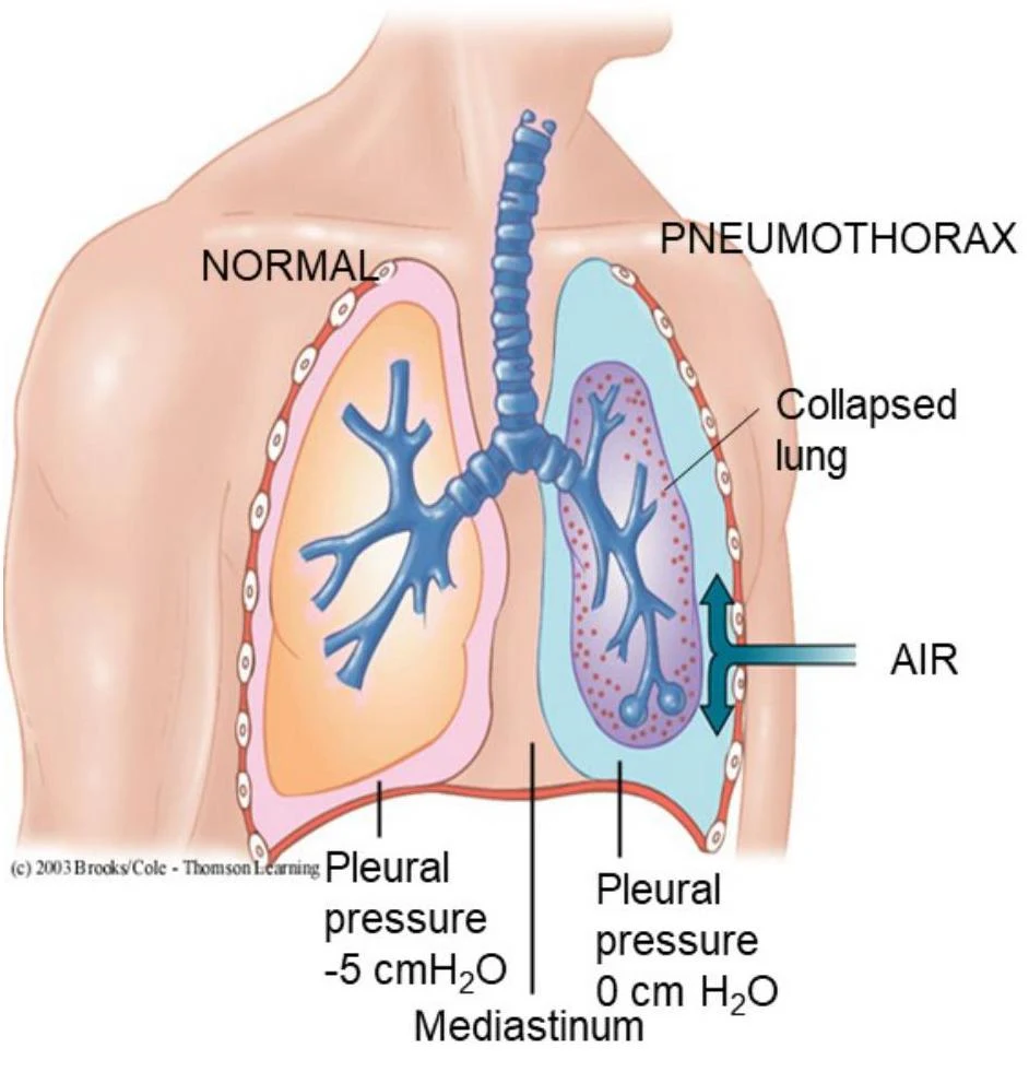
A pneumothorax occurs when air enters the intrapleural space through a breach in the chest wall or the lung. This causes the intrapleural pressure to equalize with atmospheric pressure, destroying the negative pressure gradient. Without the suction of the negative pressure, the lung's natural elastic recoil causes it to collapse.
Diagram illustrating a normal lung and a lung affected by pneumothorax, showing the difference in pleural pressure and the presence of air in the pleural space causing lung collapse.
Diagram illustrating normal lung function and the effects of a pneumothorax, alongside a chest X-ray showing a collapsed lung and mediastinal shift.
In a pneumothorax, the mediastinal membrane may prevent the opposite lung from collapsing, but it often shifts toward the unaffected side.
Keep reading for free.
Create an account to finish this reading and make it part of your study routine.