The renal and urologic systems are essential for maintaining homeostasis. By balancing the transport of solutes and water, excreting metabolic waste, and regulating acid-base balance, the kidneys ensure that the internal environment remains stable for optimal cell and tissue metabolism. Beyond simple filtration, the kidneys act as complex regulatory organs that participate in blood pressure control, red blood cell production, and bone health.
Primary Renal Functions
The kidney performs several critical tasks to maintain the body's internal equilibrium:
Homeostasis: Balancing solute and water transport to maintain stable concentrations of electrolytes and fluids.
Waste Excretion: Removing metabolic waste products from the blood.
Nutrient Conservation: Reabsorbing essential substances back into the bloodstream.
Acid-Base Regulation: Adjusting the pH of the blood by excreting acids or forming new bicarbonate ions.
Gluconeogenesis: The synthesis of glucose from amino acids.
Endocrine Functions
The kidneys are not merely filters; they are also endocrine organs that secrete vital hormones into the bloodstream:
Renin: An enzyme/hormone that plays a central role in the regulation of blood pressure via the Renin-Angiotensin-Aldosterone System (RAAS).
Erythropoietin: A hormone that stimulates the production of erythrocytes (red blood cells).
1,25-dihydroxyvitamin D3: The active form of Vitamin D, which is essential for calcium metabolism.
The Nephron: The Functional Unit
Each kidney contains approximately 1.2 million nephrons, which serve as the functional and urine-forming units of the kidney. There are three distinct types of nephrons based on their location and function:
Superficial Cortical Nephrons: These make up 85% of all nephrons and extend only partially into the medulla.
Midcortical Nephrons: These possess loops of Henle that vary in length.
Juxtamedullary Nephrons: These lie close to and extend deep into the medulla. They are critical for the process of concentrating urine and are responsible for secreting renin.
The Renal Corpuscle and Filtration
The process of urine formation begins in the renal corpuscle, which consists of the glomerulus (a tuft of capillaries) and the Bowman capsule.
The Glomerular Filtration Membrane
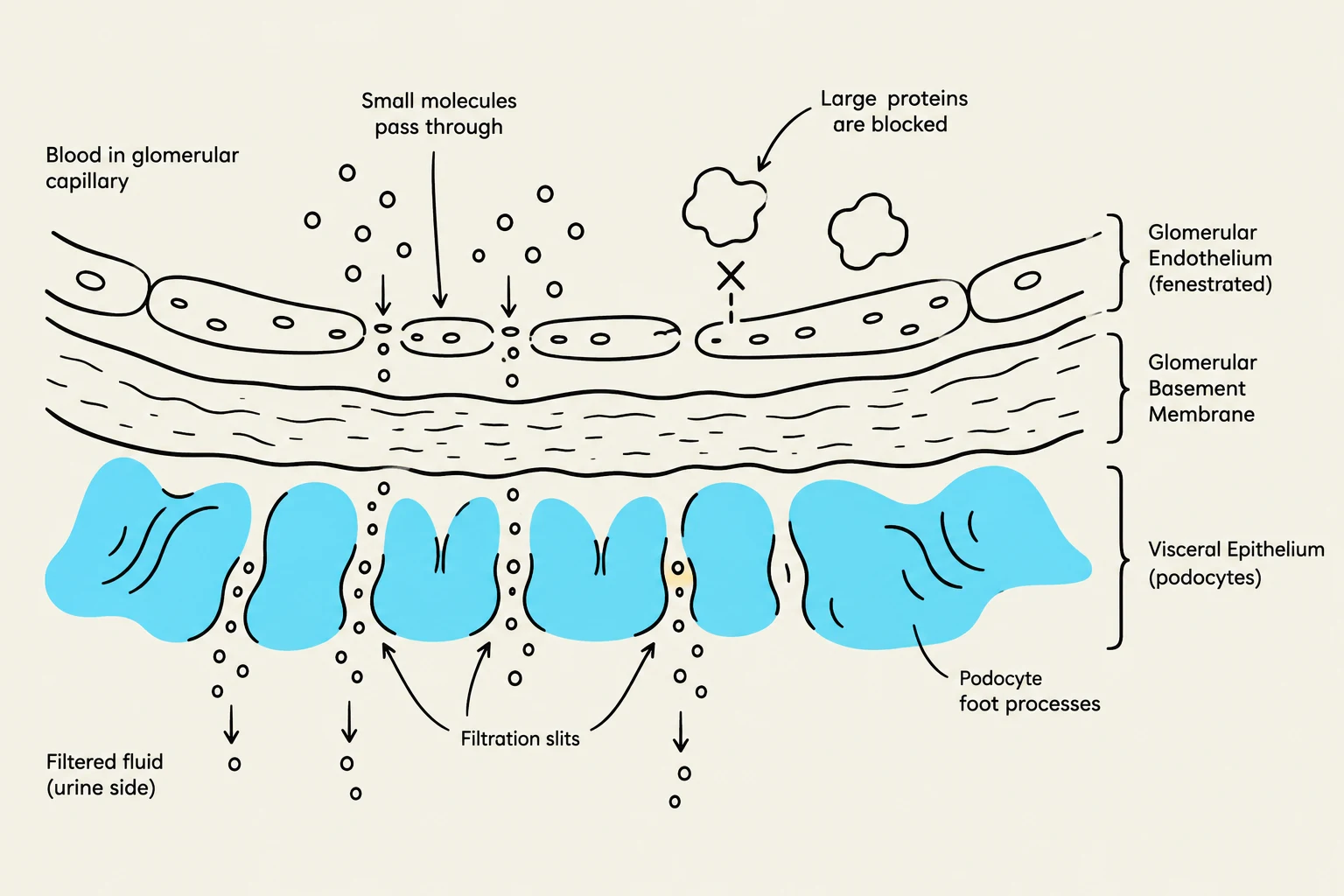
Diagram illustrating the filtration process in the glomerulus, showing how small molecules pass through the glomerular endothelium, basement membrane, and filtration slits of podocyte foot processes, while large proteins are blocked.
To form urine, plasma must pass through the glomerular filtration membrane. This membrane acts as a highly selective barrier consisting of three layers:
Glomerular Endothelium: The innermost layer, which is perforated by small openings called fenestrae. These endothelial cells also synthesize nitric oxide (a vasodilator) and endothelin-1 (a vasoconstrictor) to regulate blood flow.
Glomerular Basement Membrane (GBM): The middle layer.
Visceral Epithelium (Podocytes): The outer layer, composed of specialized cells called podocytes. These cells have footlike processes that interlace to form an elaborate network of intercellular clefts known as filtration slits.
The membrane is freely permeable to water but relatively impermeable to large colloids like plasma proteins. Permeability is influenced by both the size and the electrical charge of particles; for instance, positively charged particles permeate the membrane more easily than neutral or negative ones.
Exam Tip: Net Filtration Pressure
Remember that filtration is driven by the balance of forces. The primary force favoring filtration is the capillary hydrostatic pressure. The forces opposing filtration include the oncotic pressure within the capillary and the hydrostatic pressure within the Bowman capsule.
The Juxtaglomerular Apparatus (JGA)
The JGA is a specialized structure located where the distal tubule meets the afferent arteriole. It is essential for controlling renal blood flow (RBF), glomerular filtration rate (GFR), and renin secretion. It consists of two main components:
Juxtaglomerular Cells: Specialized cells located around the afferent arteriole.
Macula Densa: A portion of the distal convoluted tubule containing specialized cells that sense sodium and chloride levels.
The JGA facilitates tubuloglomerular feedback. For example, if sodium filtration increases, the macula densa stimulates the afferent arteriole to constrict, thereby decreasing the GFR.
Renal Tubules and Transport Mechanisms
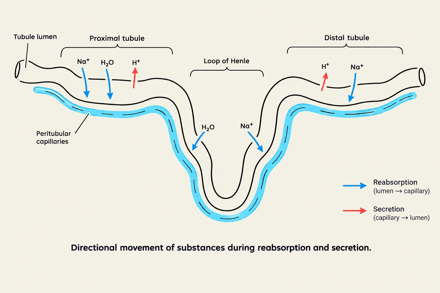
Diagram illustrating the directional movement of substances during reabsorption and secretion in the nephron, including the proximal tubule, loop of Henle, and distal tubule, with associated peritubular capillaries.
Once the filtrate enters the tubules, it undergoes three processes: filtration, reabsorption (moving substances from the lumen back to the capillaries), and secretion (moving substances from the capillaries into the lumen).
Tubular Anatomy and Function
The nephron's tubules are specialized for different transport tasks:
Segment
Primary Functions
Characteristics
Proximal Tubule
Massive reabsorption of sodium (majority) and water; secretion of hydrogen and foreign substances.
Lined with microvilli to increase surface area for reabsorption.
Loop of Henle
Transports solutes and water to maintain the hypertonic state of the renal medulla; concentrates urine.
Descending loop reabsorbs water; ascending loop reabsorbs sodium via active transport.
Distal Tubule
Adjusts acid-base balance by excreting acid and forming new bicarbonate; reabsorbs sodium and water (with ADH).
Can be isotonic or hypotonic.
Collecting Duct
Final concentration of urine; regulates sodium, potassium, and hydrogen.
Contains principal cells (sodium/water resorption) and intercalated cells (hydrogen/bicarbonate/potassium secretion).
Urine Flow Pathway
After processing in the tubules, urine follows a specific anatomical route to exit the body:
Collecting Ducts→Renal Papillae (projections of the ducts).
Minor and Major Calyces.
Renal Pelvis.
Ureters: 30 cm long smooth muscle tubes that use peristaltic activity to move urine to the bladder.
Bladder: A storage organ featuring the detrusor muscle (smooth muscle) and the trigone (a triangular area between the ureter openings and the urethra).
Urethra: The final exit pathway. In males, this is significantly longer (18–20 cm) than in females (3–4 cm).
Clinical Note: Micturition
Micturition (urination) is controlled by a reflex arc. When the bladder accumulates 250 to 300 ml of urine, mechanoreceptors sense the stretch, sending impulses to the sacral spinal cord. This triggers the contraction of the detrusor muscle and the relaxation of the internal urethral sphincter. While the internal sphincter is involuntary (autonomic), the external urethral sphincter is composed of striated skeletal muscle and is under voluntary control.
Keep reading for free.
Create an account to finish this reading and make it part of your study routine.