Structure and Function of the Cardiovascular, Peripheral Vascular, and Lymphatic Systems
A comprehensive review of the normal structures and physiological functions of the cardiovascular, peripheral vascular, and lymphatic systems, covering their roles in transport, defense, and fluid balance.
Understanding the transport and defense mechanisms of the human body requires a deep dive into three interconnected systems: the cardiovascular, peripheral vascular, and lymphatic systems. Together, these systems ensure that oxygen and nutrients reach every cell, metabolic waste is removed, fluid balance is maintained, and the immune system can respond to pathogens. This article provides a comprehensive review of their normal structures and physiological functions.
The Cardiovascular System (CVS)
The cardiovascular system is a closed circuit designed to transport blood throughout the body. The heart acts as the central pump, driving blood through two distinct but connected pathways: pulmonary circulation and systemic circulation.
In pulmonary circulation, the right side of the heart pumps deoxygenated blood to the lungs to pick up oxygen. Once oxygenated, the blood returns to the left side of the heart, which then drives systemic circulation, pumping oxygen-rich blood to the rest of the body.
Heart Anatomy and Electrical Conduction
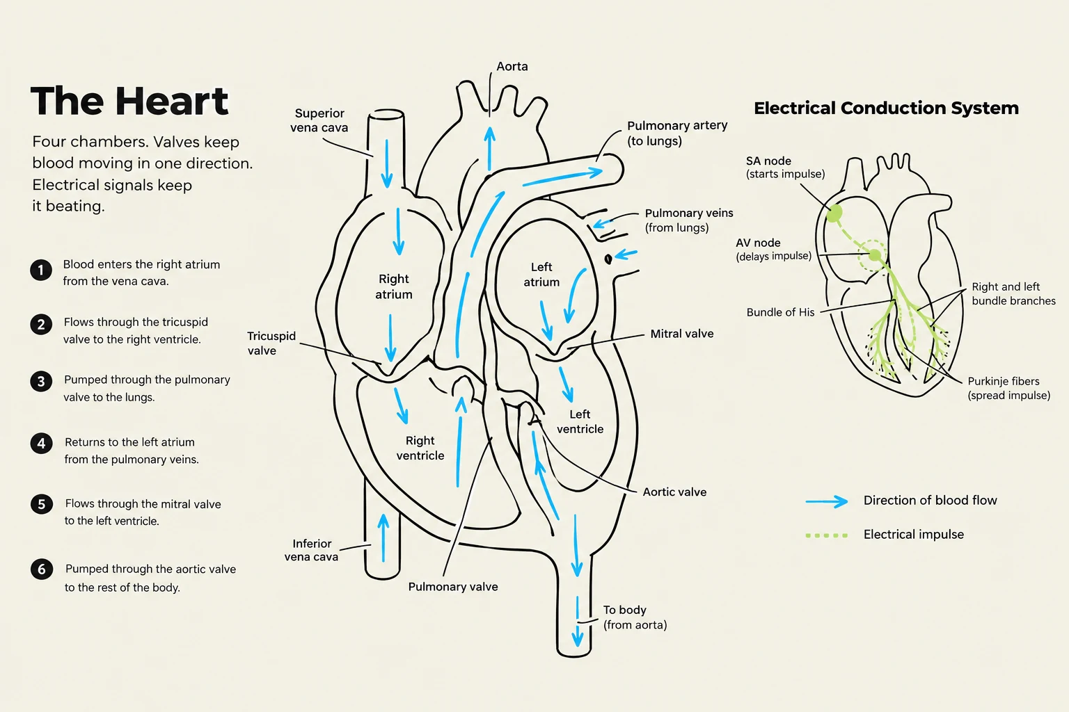
The heart is divided into four chambers: the right atrium, right ventricle, left atrium, and left ventricle. To ensure blood moves in only one direction, four valves are utilized:
Atrioventricular valves: The tricuspid (right side) and mitral (left side) valves.
Semilunar valves: The pulmonic (right side) and aortic (left side) valves.
The rhythmic contraction of the heart is governed by a specialized electrical conduction pathway. The impulse begins at the SA node (the natural pacemaker), travels to the AV node, moves through the bundle of His, down the bundle branches, and finally reaches the Purkinje fibres to trigger ventricular contraction.
Diagram illustrating the human heart's chambers, valves, and the electrical conduction system that regulates its beating, along with the direction of blood flow.
The Cardiac Cycle and Output
The cardiac cycle describes the sequence of events in a single heartbeat, divided into two phases:
Systole: The period of ventricular contraction and blood ejection.
Diastole: The period of ventricular relaxation and filling.
During auscultation (listening to the heart), clinicians hear two primary sounds. S1 ("lub") is produced by the closure of the mitral and tricuspid valves at the start of systole, while S2 ("dub") is produced by the closure of the aortic and pulmonic valves at the start of diastole.
A vital measure of how effectively the heart is meeting the body's demands is cardiac output. This is calculated using the following formula:
Cardiac Output=Heart Rate×Stroke Volume
The Peripheral Vascular System (PVS)
While the heart provides the pressure, the peripheral vascular system provides the network of vessels that distribute blood and manage pressure across the body.
Vessel Types and Function
The PVS consists of three primary types of vessels:
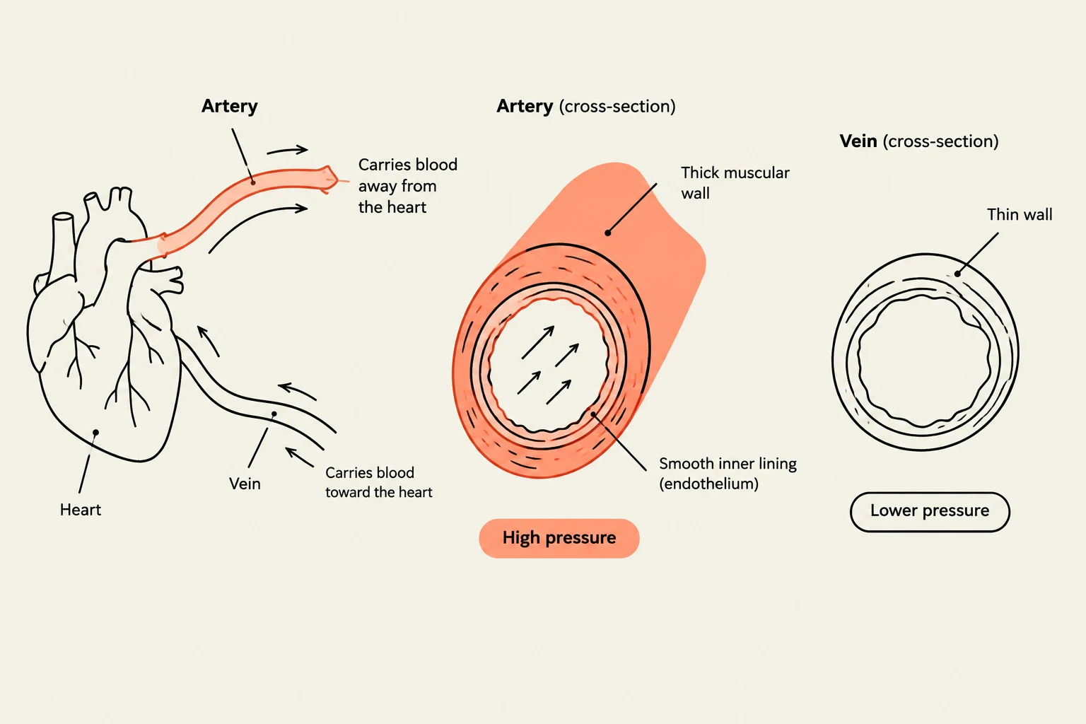
Arteries: These are high-pressure vessels with muscular, elastic walls that carry blood away from the heart. Small branches called arterioles are particularly important because they regulate vascular resistance.
Capillaries: These microscopic vessels are the functional site of the system, where the actual exchange of gases, nutrients, and fluids occurs between the blood and the tissues.
Veins: These are lower-pressure, distensible vessels that return blood to the heart.
Venous Return
Because venous pressure is low, the body employs several mechanisms to ensure venous return (the flow of blood back to the heart) against the force of gravity. Veins contain one-way valves to prevent backflow, and the calf-muscle pump—the contraction of leg muscles during movement—helps squeeze blood upward. Additionally, respiratory movements (breathing) assist in facilitating this return.
Diagram illustrating the structure of arteries and veins, showing their connection to the heart and the differences in their cross-sections and blood pressure.
The Lymphatic System
The lymphatic system serves as a secondary circulatory system that is essential for fluid homeostasis and immune defense.
Fluid Balance and Immunity
The primary role of the lymphatic vessels is to retrieve excess interstitial fluid and proteins that leak out of the capillaries and return them to the bloodstream. Without this process, significant swelling (edema) would occur. Furthermore, the lymphatic system is responsible for absorbing intestinal lipids and supporting the immune system via lymph nodes.
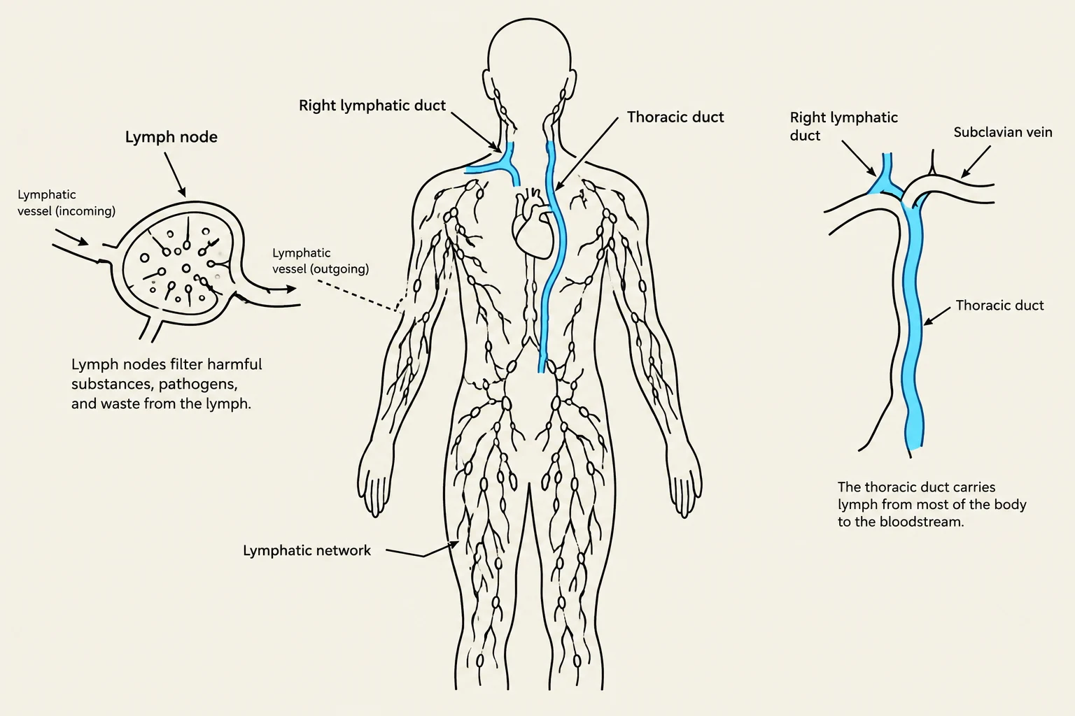
Lymph nodes act as filters, trapping pathogens and facilitating an immune response. In the head and neck region, there are ten specific groups of lymph nodes (such as submandibular, tonsillar, and cervical nodes) that drain the area into the larger lymphatic chain.
Lymphatic Drainage Pathways
Lymphatic fluid is eventually returned to the venous circulation through two major ducts:
Right lymphatic duct: Drains lymph from the right upper body.
Thoracic duct: Drains the remainder of the body.
Diagram illustrating the human lymphatic system, including lymph nodes, lymphatic vessels, the thoracic duct, and the right lymphatic duct, with a description of the thoracic duct's function.
Exam Tip: The Six P's
When assessing the peripheral vascular system, remember that the "Six Ps" are urgent warning signs of acute limb ischemia (a medical emergency):
Pain
Pallor (paleness)
Pulselessness
Paresthesia (numbness/tingling)
Paralysis
Poikilothermia (coldness of the limb).
Keep reading for free.
Create an account to finish this reading and make it part of your study routine.