


Skeletal muscle is a complex, highly organized tissue designed for a single primary purpose: movement. Whether you are lifting a heavy weight, typing on a keyboard, or simply breathing, your skeletal muscles are performing mechanical work driven by precise structural arrangements and neurological signals. To understand how a muscle moves a bone, we must look beneath the surface at the layers of connective tissue, the intricate supply of nutrients, and the electrical commands sent from the brain.
The Architecture of Muscle Tissue
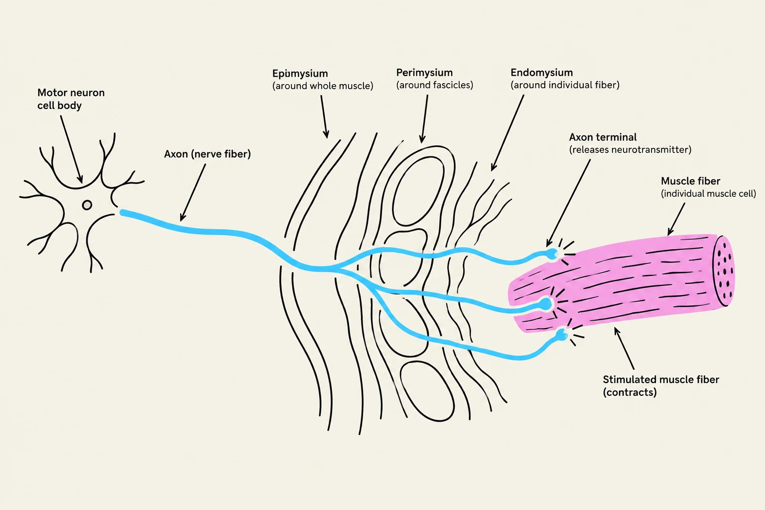
Skeletal muscle is not just a single mass of cells; it is a hierarchical structure organized by layers of connective tissue. These layers are primarily composed of collagen fibers, which provide the strength and elasticity necessary to withstand the tension of contraction.
The organization follows a specific pattern of "wrapping":
- Epimysium: The outermost layer of connective tissue that surrounds the entire muscle.
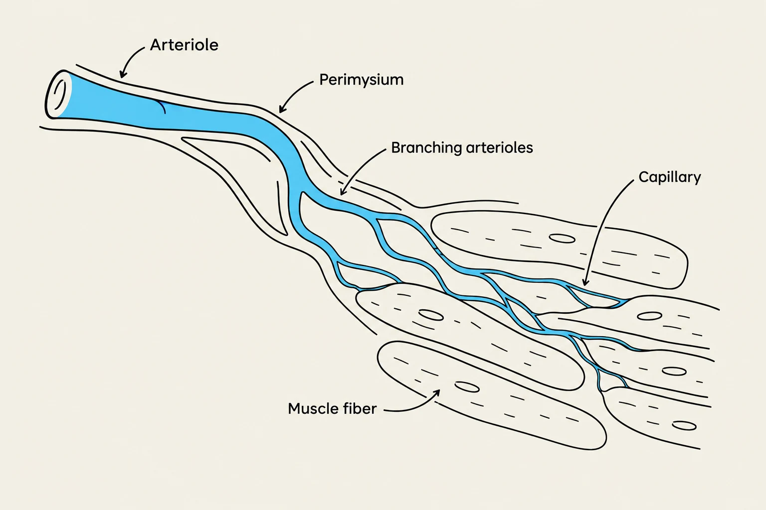
- Perimysium: A deeper layer that groups muscle fibers into bundles called fascicles.
- Endomysium: The innermost layer that surrounds each individual muscle fiber.
The collagen fibers of the perimysium and endomysium are not isolated; they are interwoven and blend into one another to create a continuous structural framework.

Attachment: Tendons and Aponeuroses
For a muscle to move a limb, it must be firmly anchored to the skeleton. At each end of a muscle, the collagen fibers from all three layers—the epimysium, perimysium, and endomysium—converge and come together. This convergence forms one of two structures:
Keep reading for free.
Create an account to finish this reading and make it part of your study routine.
Continue reading freeAlready have an account? Log in
Updated