Explores the plasma membrane's role as a selective barrier, detailing the fluid mosaic model, the lipid bilayer (phospholipids, cholesterol, glycolipids), and membrane proteins (integral, peripheral, glycoproteins). Covers selective permeability, transport mechanisms, and key cellular functions.
The survival of a cell depends entirely on its ability to maintain a distinct internal environment that is different from the world outside. This boundary is provided by the plasma membrane, a flexible yet sturdy barrier that surrounds and contains the cytoplasm of a cell. By separating the cytosol—the fluid component of the cytoplasm—from the surrounding extracellular fluid, the plasma membrane allows the cell to regulate its internal chemistry and respond to external stimuli.
The Fluid Mosaic Model
To understand how such a thin barrier can be both strong and flexible, scientists use the fluid mosaic model. This model describes the membrane not as a static wall, but as a continually moving "sea" of fluid lipids.
Within this lipid sea, various proteins are embedded, creating a "mosaic" pattern. Some proteins float freely like icebergs in the ocean, while others are anchored firmly in place like islands. This fluidity is essential, as it allows molecules to move laterally within the membrane, facilitating processes like cell signaling and the movement of proteins to where they are needed.
Figure 3.2: The fluid mosaic arrangement of lipids and proteins in the plasma membrane.
The Lipid Bilayer: The Structural Foundation
The fundamental framework of the membrane is the lipid bilayer, which consists of two back-to-back layers of lipid molecules. This bilayer is composed of three primary types of lipids, each serving a unique role in maintaining the membrane's integrity:
1. Phospholipids
Accounting for approximately 75% of the membrane lipids, phospholipids are the most abundant component. They are "amphipathic" molecules, meaning they have two distinct personalities:
Hydrophilic Heads: The circular "heads" contain phosphorus and are "water-loving," meaning they face outward toward the aqueous extracellular fluid and the internal cytosol.
Hydrophobic Tails: The two long, wavy fatty acid tails are "water-fearing." They face inward, away from the water, creating a nonpolar, oily core in the center of the membrane.
2. Cholesterol
Comprising about 20% of the lipids, cholesterol molecules are interspersed between the phospholipid tails. These steroid molecules act as "temperature buffers," providing stability to the membrane and preventing it from becoming too fluid in heat or too rigid in cold.
3. Glycolipids
Making up about 5% of the lipids, glycolipids are lipids that have carbohydrate groups attached to them. These molecules extend from the membrane surface into the extracellular space.
Study Tip: Remember that the "hydrophobic core" created by the phospholipid tails is the reason why many substances cannot simply pass through the membrane. It acts as a chemical barrier to anything that is not lipid-soluble.
Membrane Proteins: The Functional Machinery
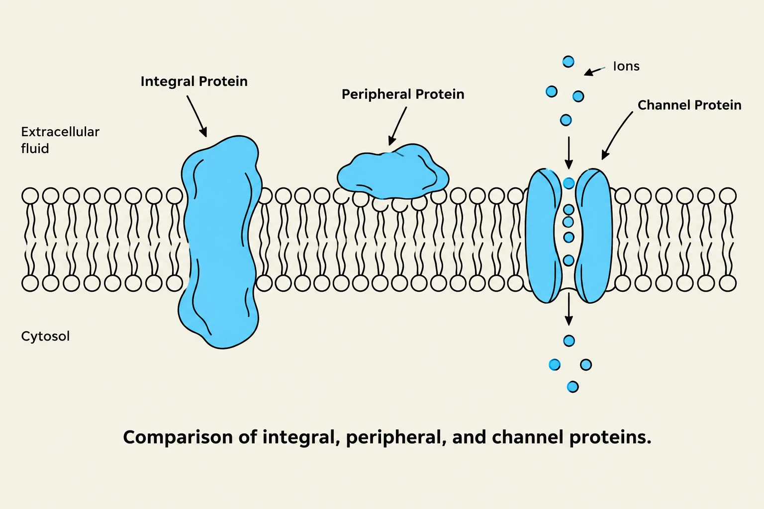
Comparison of integral, peripheral, and channel proteins in a cell membrane.
While the lipids provide the structure, the proteins embedded within the bilayer provide the function. Proteins are categorized based on their position within the membrane:
Integral (Transmembrane) Proteins: These are large, complex structures that span the entire width of the lipid bilayer, from the extracellular fluid to the cytosol. A vital subtype is the channel protein (or pore), which creates a water-filled tunnel through the hydrophobic core, allowing polar molecules and ions to pass through.
Peripheral Proteins: These are smaller structures located on the inner or outer surfaces of the membrane. They do not span the bilayer but often attach to integral proteins or the lipid heads to assist in signaling or maintaining the cell's shape.
Furthermore, many proteins are modified with carbohydrates to become glycoproteins. Together with glycolipids, these carbohydrate-attached molecules serve as "ID tags" for the cell, allowing the immune system to recognize the cell as "self" rather than a foreign invader.
Selective Permeability and Transport
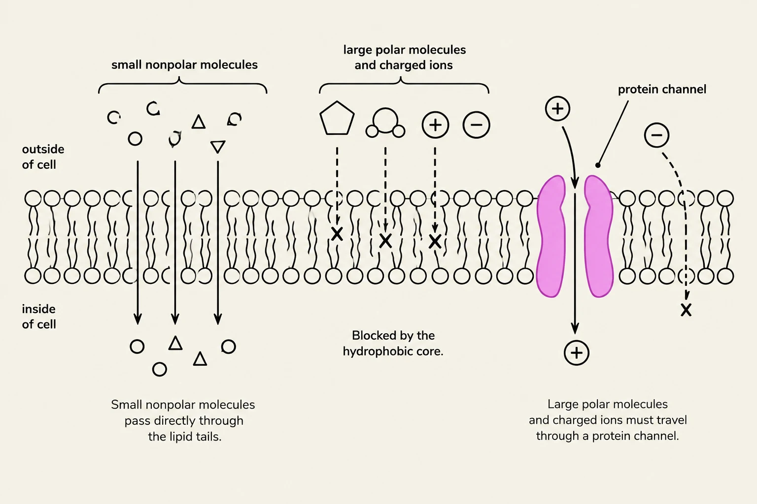
This diagram illustrates how different types of molecules cross the cell membrane. Small nonpolar molecules can pass directly through the lipid bilayer, while large polar molecules and charged ions require protein channels for transport.
The plasma membrane is not equally open to all substances; it exhibits selective permeability. This means it acts as a gatekeeper, allowing some substances to pass easily while blocking others.
What passes easily? Lipid-soluble (nonpolar) molecules can dissolve through the hydrophobic core of the bilayer.
What is blocked? Charged ions (like Na+ or $K^+$) and large polar molecules cannot cross the oily interior of the membrane on their own.
To move these blocked substances, the cell relies on integral proteins like channel proteins. The movement of these ions is often driven by an electrochemical gradient—a combination of the concentration gradient (the difference in substance concentration) and the electrical gradient (the difference in charge across the membrane).
Summary of Functions
To wrap up, the plasma membrane serves four critical roles in cellular biology:
Physical Barrier: It separates the internal cytosol from the external environment.
Regulated Transport: It controls the flow of substances into and out of the cell via selective permeability.
Cell Identification: Through glycoproteins and glycolipids, it helps cells recognize one another.
Intercellular Signaling: It facilitates communication between cells, allowing them to work together as part of a larger organism.
Keep reading for free.
Create an account to finish this reading and make it part of your study routine.