Explore the dynamic structure of the plasma membrane, its lipid bilayer composition, and the crucial roles of membrane proteins in cellular functions like transport, signaling, and enzymatic activity. Learn about passive and active transport mechanisms and the importance of compartmentalization in eukaryotic cells.
The plasma membrane is far more than a simple boundary; it is a dynamic, complex, and asymmetric structure that defines the very essence of a cell. In eukaryotic cells, this membrane serves as the primary interface between the internal environment and the outside world, managing everything from nutrient uptake to signal detection. While the membrane provides a fundamental permeability barrier, it is the intricate arrangement of lipids and proteins that allows the cell to maintain homeostasis and perform specialized biological tasks.
The Lipid Bilayer: The Fundamental Barrier
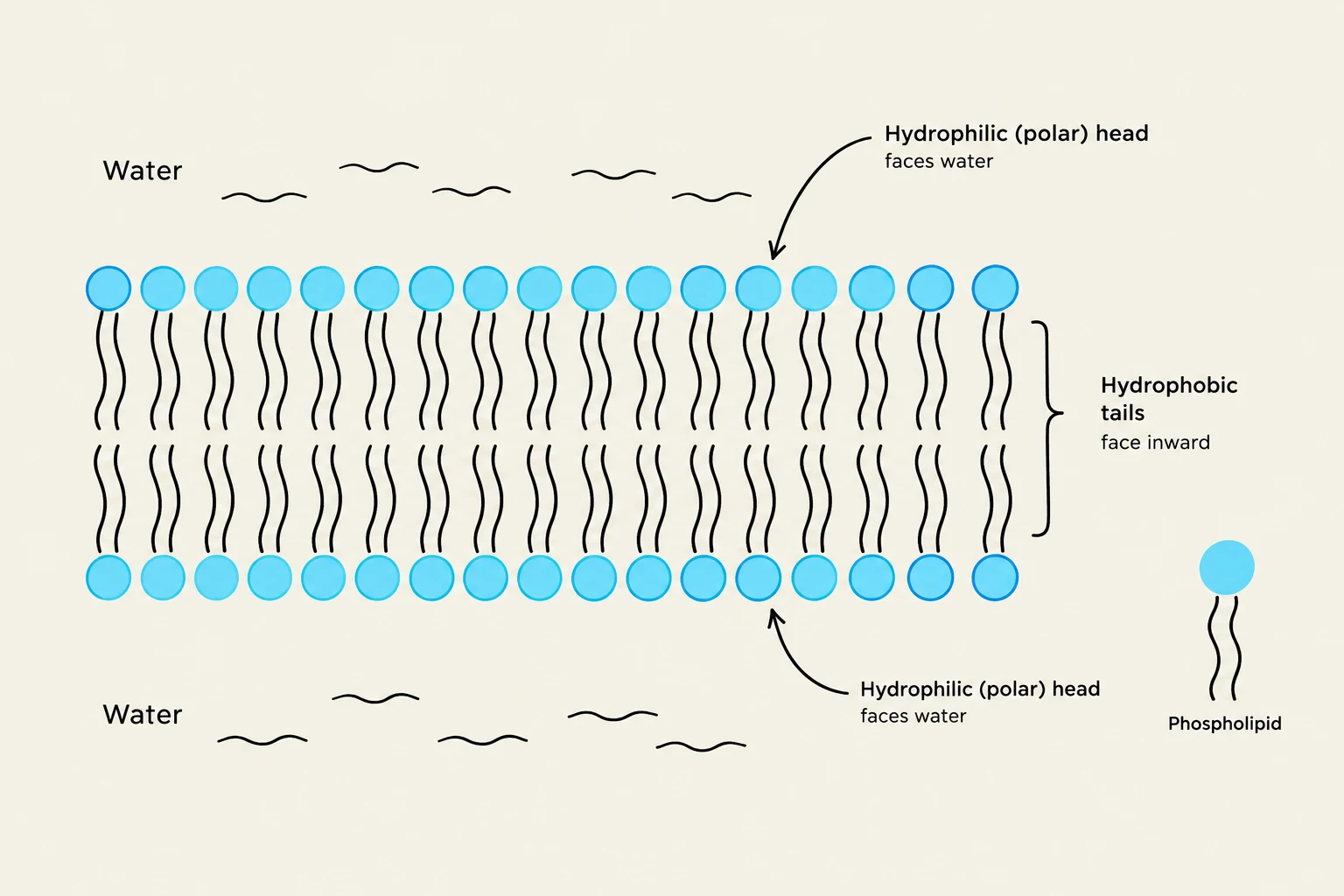
At the core of every cell membrane is the lipid bilayer. This structure is driven by the behavior of amphipathic molecules—lipids that possess both a hydrophilic ("water-loving") head and a hydrophobic ("water-fearing") tail. In an aqueous environment, these molecules spontaneously organize into a bilayer to minimize the energy cost of exposing hydrophobic regions to water. The hydrophilic heads face the water on both the extracellular and cytosolic sides, while the hydrophobic tails are shielded within the interior, much like the filling in a sandwich.
The bilayer is composed of several key lipid types, each contributing to the membrane's unique properties:
Phospholipids: The most abundant lipids, consisting of a phosphate-containing head and two hydrocarbon tails. They exhibit asymmetric distribution; for instance, phosphatidylserine and phosphatidylethanolamine are primarily found on the cytosolic side, while phosphatidylinositols are concentrated there to assist in cell signaling.
Glycolipids: These contain sugar groups and are found exclusively in the noncytosolic (extracellular) monolayer, often serving in cell recognition.
Cholesterol: A sterol molecule distributed relatively equally between both monolayers, helping to regulate membrane fluidity.
Diagram illustrating the structure of a phospholipid bilayer, showing hydrophilic heads facing water and hydrophobic tails facing inward.
Exam Tip: Remember that the bilayer is "self-sealing." Because exposing hydrophobic tails to water is energetically unfavorable, any small tear in the membrane will spontaneously rearrange to close the gap.
Membrane Fluidity and Composition
The lipid bilayer is not a static wall; it is a two-dimensional fluid. Individual lipid molecules can move rapidly within the plane of their own monolayer through lateral diffusion. However, "flip-flopping"—moving from one monolayer to the other—is extremely rare because it requires the hydrophilic head to pass through the hydrophobic core.
The ease of this movement, or fluidity, depends heavily on the chemical composition of the tails:
Tail Length: Shorter hydrocarbon chains reduce van der Waals interactions, increasing fluidity.
Saturation: The presence of double bonds (unsaturation) creates "kinks" in the tails, preventing tight packing and thus increasing fluidity.
Membrane Proteins: The Functional Engines
While lipids provide the structure, membrane proteins execute the cell's specialized functions. These proteins are often transmembrane proteins, meaning they span the entire bilayer multiple times. They can be categorized by their primary roles:
Transporters: These move specific solutes across the membrane. They are divided into channels (which form pores for rapid movement) and transporters (which undergo conformational changes to move molecules).
Receptors: These detect extracellular signals (like hormones) and trigger internal cellular responses.
Enzymes: Proteins that catalyze specific chemical reactions at the membrane surface.
Anchors: These tether the membrane to the internal cytoskeleton or the external extracellular matrix, providing structural stability.
Diagrams illustrating the difference between ion channel proteins and transporter proteins in cell membranes.
Principles of Transmembrane Transport
The hydrophobic interior of the bilayer creates a significant barrier to most hydrophilic molecules. The rate at which a molecule can cross via simple diffusion depends on its size and solubility:
Small, uncharged polar molecules (H2O, ethanol): Diffuse at a modest rate.
Large uncharged polar molecules (glucose): Cross very slowly.
Ions (Na+, $K^+$, $Cl^-$): Highly impermeable to the bilayer regardless of size.
To overcome this barrier, cells use facilitated transport via specialized proteins.
Passive vs. Active Transport
Transport mechanisms are classified by their energy requirements:
Passive Transport: Movement "downhill" from high to low concentration. For uncharged molecules, this is driven by the concentration gradient. For charged ions, the direction is determined by the electrochemical gradient, which is the sum of the concentration gradient and the membrane potential (the voltage difference across the membrane).
Osmosis: A specific type of passive transport where water moves down its concentration gradient. Cells often use aquaporins (specialized water channels) to accelerate this process.
Active Transport: Movement "uphill" against a concentration gradient, which requires an input of energy. This is performed by pumps:
ATP-driven pumps: Use energy from ATP hydrolysis (e.g., the Na+-$K^+$ ATPase).
Gradient-driven pumps: Couple the downhill movement of one solute to the uphill movement of another.
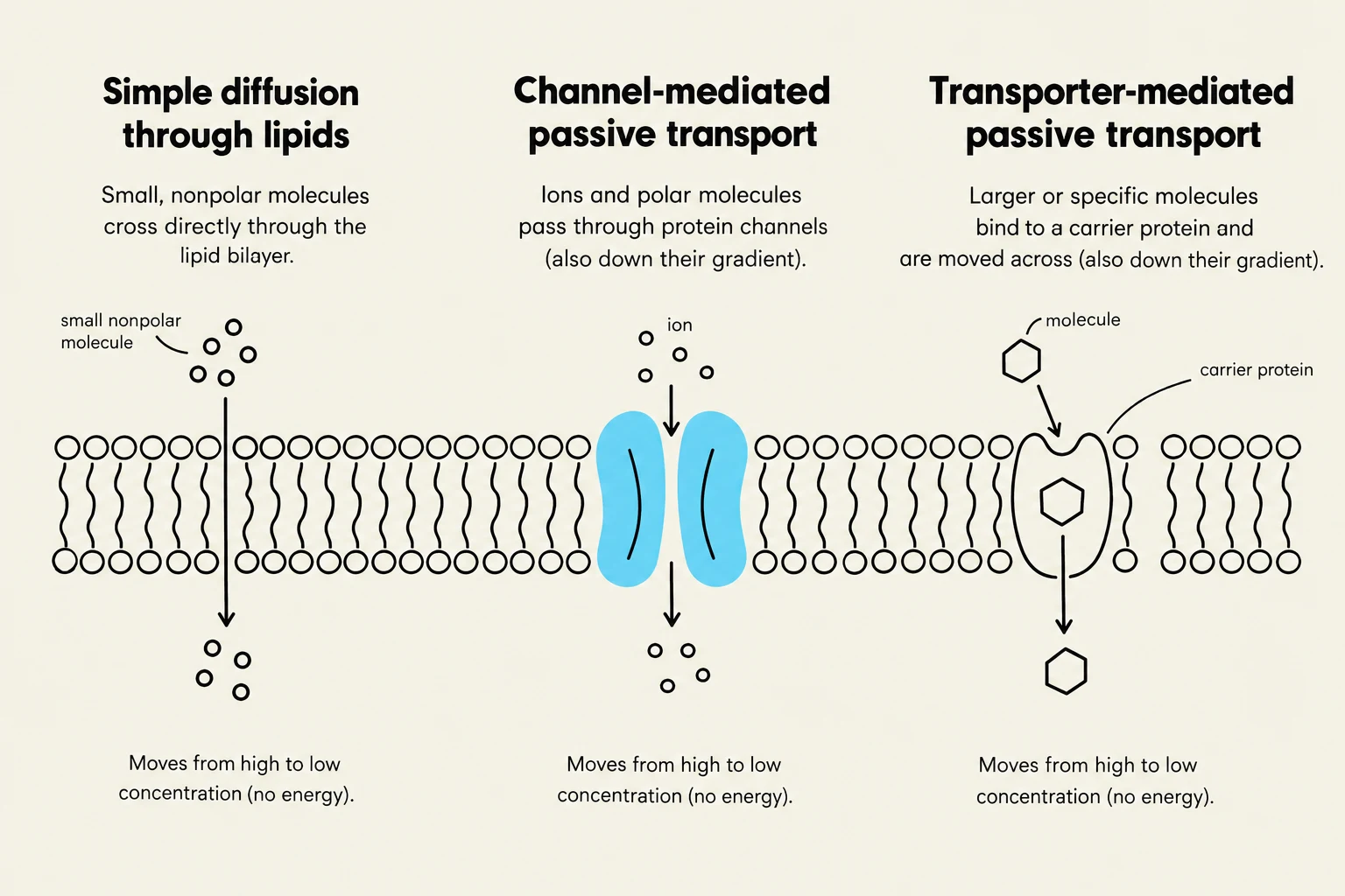
Diagrams illustrating three types of passive transport across a cell membrane: simple diffusion through lipids, channel-mediated passive transport, and transporter-mediated passive transport. All processes move substances from high to low concentration without energy expenditure.
Compartmentalization and Organelles
In eukaryotic cells, the plasma membrane is just one part of an elaborate system of internal membranes. This compartmentalization allows the cell to segregate distinct metabolic processes, a necessity for cells that are 1,000 to 10,000 times larger in volume than prokaryotes.
Major membrane-enclosed organelles include:
Nucleus: Enclosed by a double membrane (the nuclear envelope) and contains the genome.
Endoplasmic Reticulum (ER): The site of lipid and protein synthesis. The Rough ER is studded with ribosomes for protein synthesis, while the Smooth ER handles lipid synthesis and detoxification.
Golgi Apparatus: Modifies, sorts, and packages proteins and lipids for delivery.
Lysosomes: Contain digestive enzymes to break down macromolecules and worn-out organelles.
Mitochondria: The site of ATP synthesis via oxidative phosphorylation.
These organelles are not floating aimlessly; they are positioned and moved by the cytoskeleton (primarily microtubules) using motor proteins that consume ATP. This complex internal architecture allows the eukaryotic cell to function as a highly organized, efficient biological machine.
Keep reading for free.
Create an account to finish this reading and make it part of your study routine.