This content examines the structure and function of the nucleus, ribosomes, rough endoplasmic reticulum, Golgi apparatus, and the secretory pathway. It also covers protein folding, modification, degradation, and the roles of proteasomes and lysosomes.
Cells are the basic units of life, enclosed by a lipid bilayer membrane and containing a complex internal environment known as the cytoplasm. This cytoplasm is composed of the cytosol and various specialized structures called organelles. To understand how a cell functions—for example, how a pancreatic cell produces and releases insulin to regulate blood sugar—one must understand the intricate relationship between these organelles and the pathways they form.
The Command Center: The Nucleus
The nucleus serves as the primary control center of the cell, housing the genetic material required for instruction and replication. Within the nucleus, DNA is organized into a nucleoprotein complex known as chromatin, which consists of DNA associated with histone proteins and non-histone proteins.
Chromatin exists in two distinct structural states that dictate gene activity:
Euchromatin: This is a loosely arranged, less condensed form of chromatin that is transcriptionally active, allowing the cellular machinery to access the DNA for gene expression.
Heterochromatin: This is highly condensed and transcriptionally inactive, meaning the genes within these regions are not being expressed.
The nucleus is not a single open space; it contains the nucleoplasm, a matrix that acts as an organizational scaffold and contains various nuclear particles, such as protein/RNA complexes involved in processing pre-mRNA. A prominent feature within the nucleus is the nucleolus, a non-membrane-bound inclusion most visible in cells actively synthesizing proteins. The nucleolus is the site of ribosomal RNA (rRNA) synthesis and the initial assembly of ribosomes.
Diagram illustrating the structure of a cell nucleus, including the nuclear envelope, nucleolus, nuclear pores, euchromatin, and heterochromatin, along with a flowchart explaining how these components interact in gene expression.
The nucleus is enclosed by the nuclear envelope, a double membrane structure. The outer nuclear membrane and the perinuclear cisternae are continuous with the rough endoplasmic reticulum (rER). To allow for the exchange of materials between the nucleus and the cytoplasm, the envelope is interrupted by nuclear pores, which mediate the translocation of molecules. Supporting the inner nuclear membrane is the nuclear lamina, a structure that provides mechanical stability.
Protein Synthesis: Ribosomes and the Rough ER
Once genetic instructions are transcribed, the process of protein synthesis begins. This is carried out by ribosomes, which are not membrane-enclosed but are instead RNA-protein complexes composed of small (40S) and large (60S) subunits. Ribosomes can be found free in the cytoplasm or bound to the endoplasmic reticulum. When multiple ribosomes are attached to a single mRNA strand, the structure is called a polysome.
The Rough Endoplasmic Reticulum (rER) is a system of interconnected sacs called cisternae, studded with ribosomes, giving it a "rough" appearance. The rER is particularly prominent in cells specialized for secreting proteins.
The destination of a protein is determined by specific signal sequences. Translation typically begins on a ribosome in the cytosol. If the growing polypeptide contains an rER signal peptide, translation continues on the rER membrane. The rER is responsible for synthesizing and processing proteins destined for the Golgi apparatus, lysosomes, membranes, and secretion from the cell.
Protein Folding and Modification in the rER
Inside the rER, proteins undergo critical post-translational modifications to ensure they are functional. One of the most common modifications is N-linked glycosylation, where an oligosaccharide is covalently added to the side chain of an Asparagine (Asn/N) residue. This process is vital because it helps prevent protein aggregation and aids in the binding of the protein to molecular chaperones.
Chaperones are specialized proteins that assist in the folding of nascent (newly forming) polypeptides. They bind to the polypeptide to ensure it reaches its correct three-dimensional shape and can bind to incorrectly folded proteins to prevent them from aggregating. Some chaperones, such as heat-shock proteins (Hsp), are expressed at high temperatures to help prevent protein denaturation.
Exam Tip: If a question asks which activity is occurring when a polypeptide is bound by a chaperone in the rER, remember that chaperones are primarily concerned with protein folding and preventing misfolding or aggregation.
The Secretory Pathway and the Golgi Apparatus
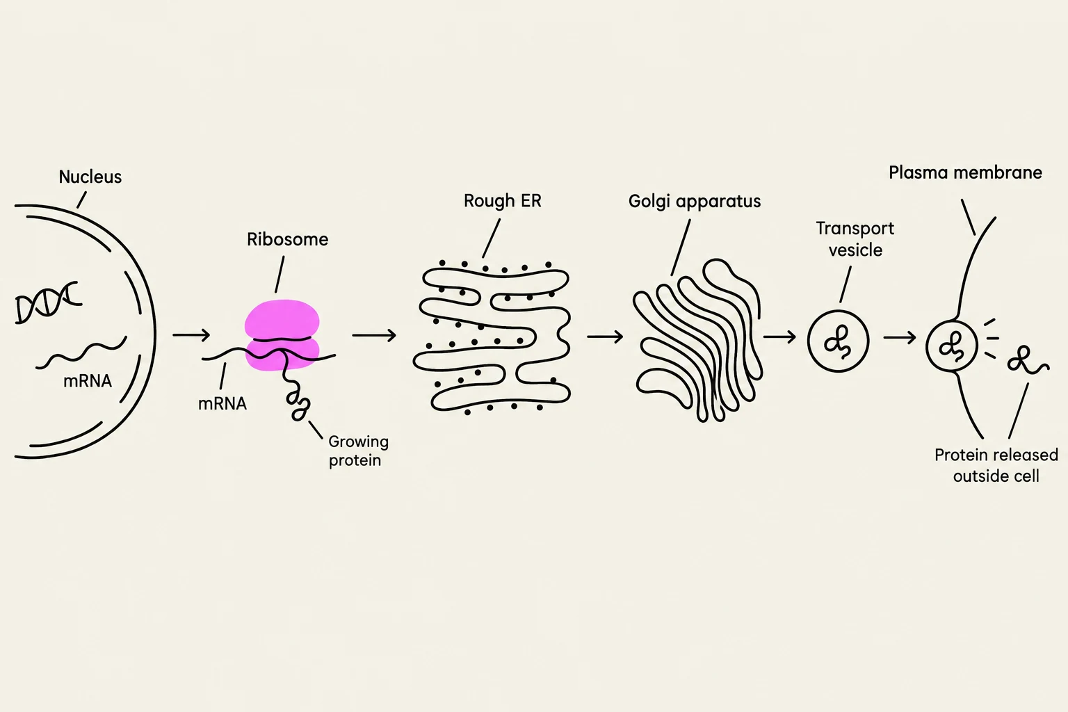
Proteins that are destined for secretion, such as insulin in pancreatic β cells, follow a specific route known as the secretory pathway.
rER: Synthesis, N-linked glycosylation, and folding occur here.
Transport Vesicles: Properly folded proteins are packaged into vesicles and sent to the Golgi apparatus via anterograde transport.
Golgi Apparatus: This organelle consists of a series of stacked, flattened cisternae. It acts as a sorting and packaging center.
The Golgi apparatus performs several critical functions:
Completing N-linked glycosylation: It refines the sugar chains added in the rER.
O-linked glycosylation: It facilitates the addition of carbohydrates to the oxygen atom of Serine or Threonine residues, which helps maintain protein stability and is essential for cell recognition and adhesion.
Sorting: The cis-Golgi network receives proteins, while the trans-Golgi network sorts them into vesicles for their final destinations.
Diagram illustrating the process of protein synthesis and secretion from a cell, starting with DNA in the nucleus, transcription to mRNA, translation by ribosomes, modification in the rough ER and Golgi apparatus, packaging into transport vesicles, and finally secretion outside the plasma membrane.
The final step of this pathway is exocytosis, the process by which transport vesicles fuse with the plasma membrane to release their contents into the extracellular environment.
Summary of the Secretory Pathway
Stage
Organelle
Key Process
1
Nucleus
Transcription of DNA to mRNA
2
Ribosome / rER
Translation and N-linked glycosylation
3
Golgi Apparatus
O-linked glycosylation, sorting, and packaging
4
Plasma Membrane
Exocytosis (release of protein)
Degradation and Quality Control
Not all proteins are destined for secretion. Cells must also manage damaged, misfolded, or unnecessary proteins through degradation pathways.
The Proteasome
Misfolded or damaged proteins (including those damaged by Reactive Oxygen Species) are often targeted for destruction by the ubiquitin-proteasome system. Proteins destined for this pathway are first "tagged" with a small protein called ubiquitin (polyubiquitination). The proteasome is a non-membrane-bound protein complex located predominantly in the cytosol that enzymatically degrades these tagged proteins.
The Lysosome
For larger cellular structures or extracellular particles brought in via phagocytosis, the cell utilizes the lysosome. Lysosomes are organelles that perform enzymatic degradation through the action of acid hydrolases in an acidified luminal space.
A critical aspect of lysosomal function is the "destination tag." Lysosomal enzymes are synthesized in the rER and must receive a mannose 6-phosphate (M6P) tag in the Golgi apparatus to be correctly directed to the lysosome.
Clinical Correlation: In I-cell disease (Mucolipidosis type II), there is a genetic error in the post-translational modification required to add the M6P tag. Consequently, lysosomal enzymes are not sent to the lysosomes, leading to the accumulation of undigested "inclusion bodies" within the cells, which causes severe neurological and physical deterioration.
Keep reading for free.
Create an account to finish this reading and make it part of your study routine.