


The vascular system is a complex network of vessels designed to transport blood throughout the entire body, ensuring that every cell receives nutrients and oxygen while effectively removing metabolic waste. This system is organized into two primary circuits: the systemic circulation, which extends to all body regions, and the pulmonary circulation, which consists of the vessels traveling to and from the lungs.
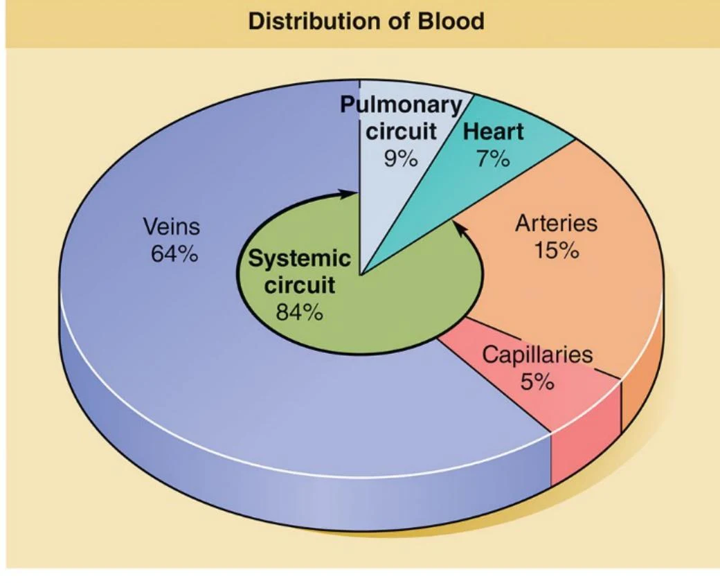
The Three Main Vessel Types
The vascular system is categorized into three distinct types of vessels based on their structure and direction of blood flow: arteries, capillaries, and veins.
Arteries
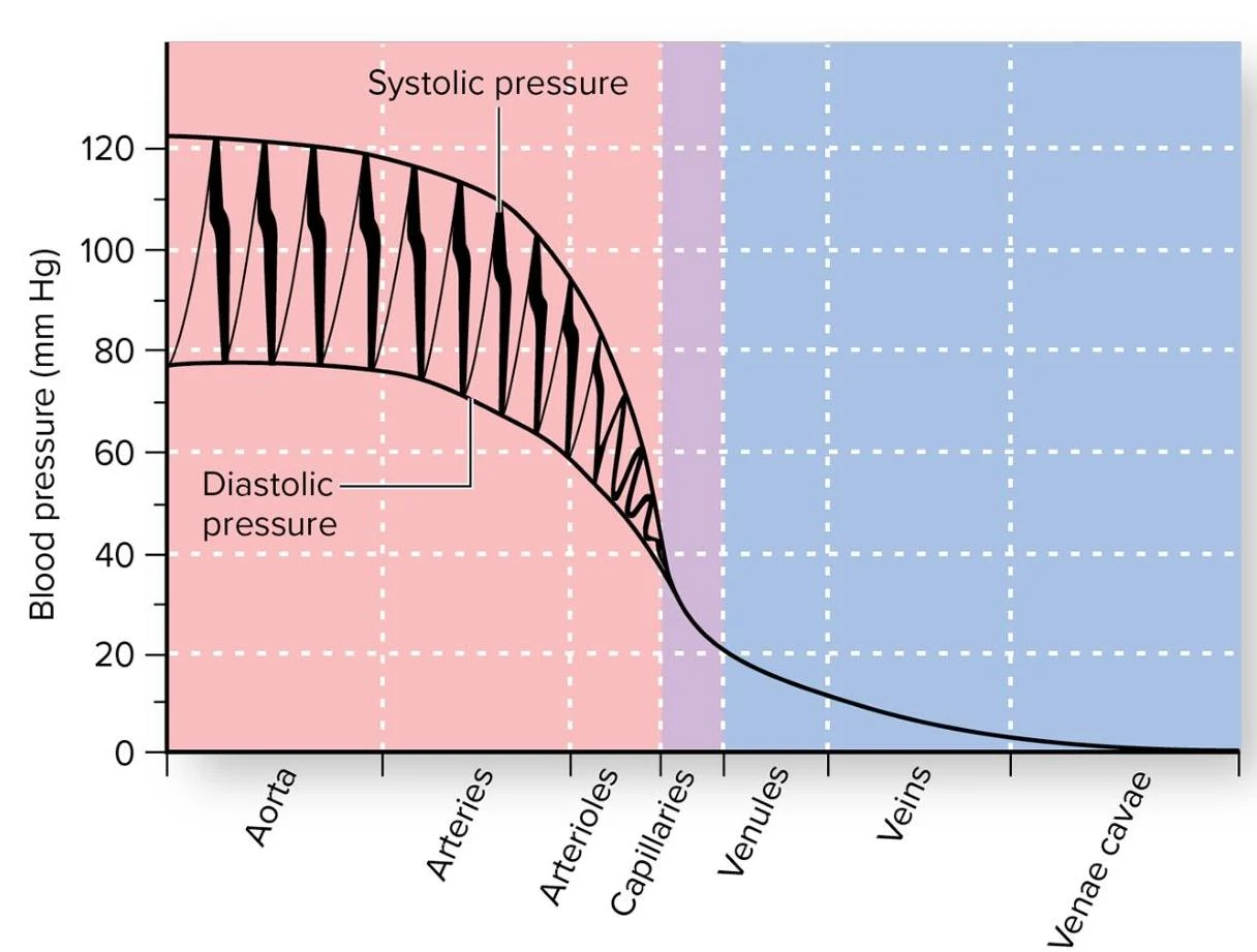
Arteries are "resistance vessels" because they must withstand high blood pressure. Their primary role is to carry blood away from the heart. As arteries move further from the heart, they branch into progressively smaller vessels.
Capillaries
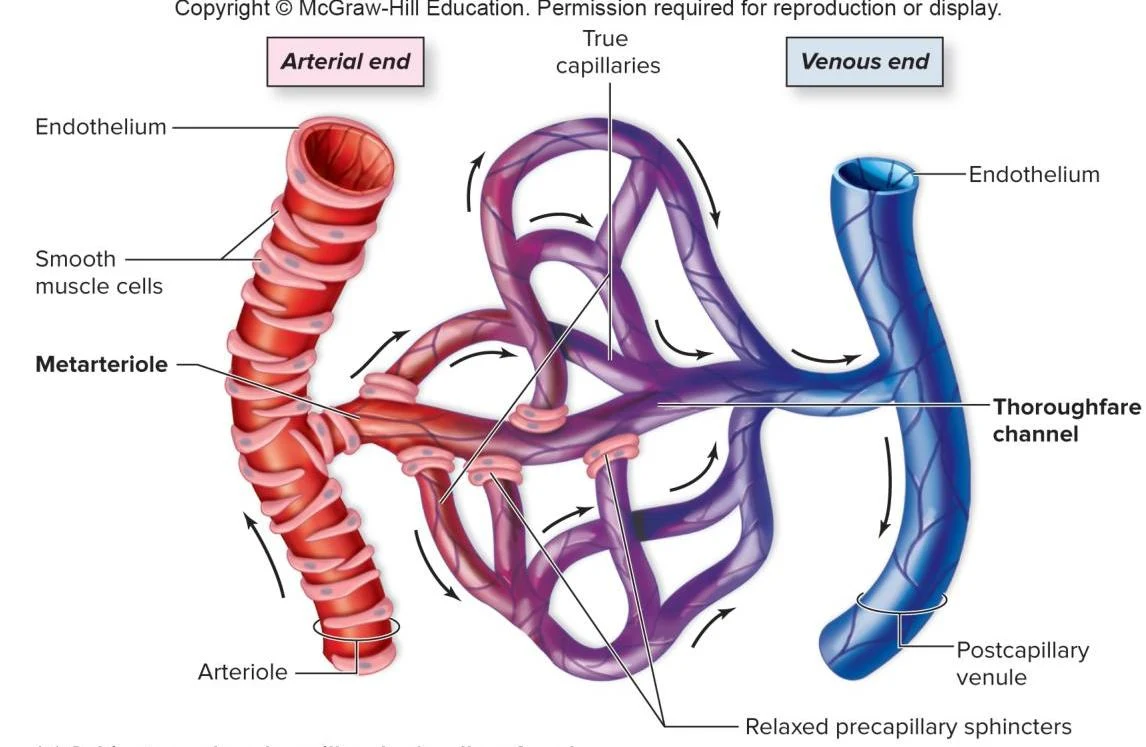
Capillaries are microscopic vessels that connect small arteries to small veins. They serve as "exchange vessels," where the actual transfer of nutrients, wastes, and hormones occurs between the blood and the surrounding tissue fluid.
Veins
Veins are "capacitance vessels," meaning they hold the majority of the body's blood volume. They function to return blood to the heart. Unlike arteries, veins become progressively larger as they merge and approach the heart.
Histology: The Three Tunics
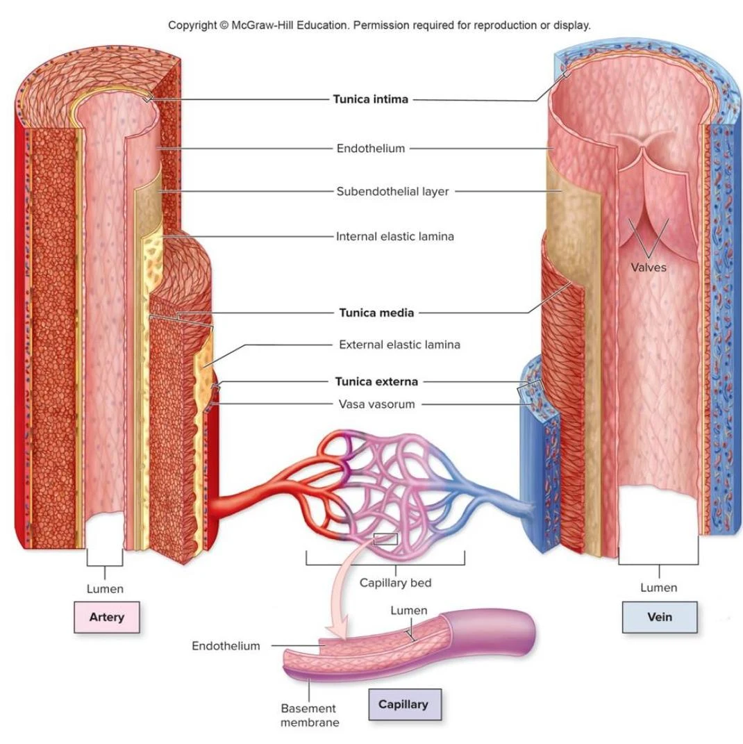
Most blood vessels (with the exception of capillaries) are composed of three distinct layers, known as tunics.
- Tunica interna (intima): The innermost layer, consisting of a simple squamous endothelium. This provides a slick, selectively permeable barrier for blood flow.
- Tunica media: The middle layer, which is typically thick and composed of smooth muscle, collagen, and sometimes elastin. This layer is responsible for vasoconstriction (narrowing of the vessel) and vasodilation (widening of the vessel).
- Tunica externa (adventitia): The outermost layer, made of loose connective tissue that anchors the vessel to its surrounding structures.


Keep reading for free.
Create an account to finish this reading and make it part of your study routine.
Continue reading freeAlready have an account? Log in
Updated