Structure and Contraction of Skeletal Muscle Fibers
Explores the microscopic structure of skeletal muscle fibers, including myofibrils, myofilaments (actin, myosin, tropomyosin, troponin, titin), and their organization into sarcomeres. Details the sliding filament mechanism of muscle contraction and the role of dystrophin in force transfer, with a clinical correlation t
Skeletal muscle is a highly organized tissue designed for voluntary movement, characterized by its ability to contract and shorten when stimulated. At the microscopic level, a skeletal muscle fiber (also known as a muscle cell) is a complex structure packed with specialized proteins that allow it to generate force. The secret to this force lies in the precise arrangement of protein filaments called myofilaments, which are organized into repeating units that create the visible stripes, or striations, seen under a microscope.
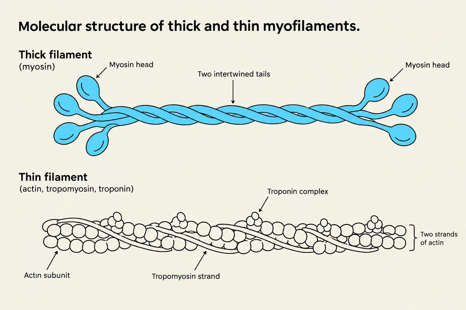
Diagram illustrating the molecular structure of thick and thin myofilaments, showing myosin heads, intertwined tails, actin subunits, tropomyosin strands, and troponin complexes.
The Building Blocks: Myofibrils and Myofilaments
The cytoplasm of a muscle fiber, known as the sarcoplasm, is occupied by long, proteinaceous cords called myofibrils. These myofibrils are the actual contractile elements of the cell. Within each myofibril, there are three distinct types of myofilaments that work in concert to facilitate movement: thick filaments, thin filaments, and elastic filaments.
Thick Filaments and Myosin
The thick filaments are composed primarily of a motor protein called myosin. Each myosin molecule is shaped somewhat like a golf club: it features two intertwined chains that form a shaft-like tail and two globular heads. These heads are arranged in a helical array around the filament, with some angling to the left and others to the right. The primary function of these myosin heads is to bind to actin, acting as the "motors" that pull the muscle into a contracted state.
Thin Filaments: Actin, Tropomyosin, and Troponin
Thin filaments are more complex, consisting of three different protein types that regulate the contraction process:
Fibrous (F) actin: This forms the structural backbone of the thin filament. It consists of two intertwined strands of globular (G) actin subunits, each containing an active site where a myosin head can attach.
Tropomyosin: This protein acts as a physical barrier. It wraps around the actin strands, blocking the active sites and preventing myosin from binding when the muscle is at rest.
Troponin: A small, calcium-binding protein located on each tropomyosin molecule. It acts as the molecular "switch" for contraction.
Elastic Filaments and Titin
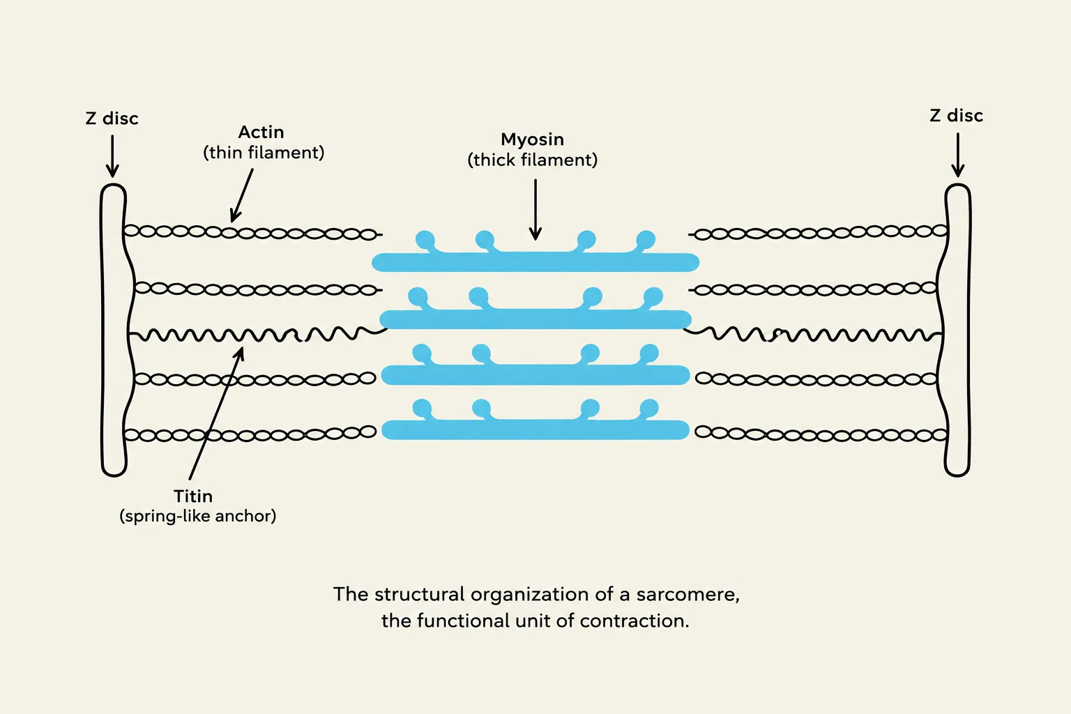
To maintain structural integrity, muscle fibers utilize elastic filaments made of a massive, springy protein called titin. Titin runs through the core of the thin filaments and anchors them to both the Z disc and the M line. This provides stability, helps position the thick filaments correctly, and prevents the muscle from being overstretched.
Study Tip: Protein Categorization
On exams, you may be asked to categorize these proteins by function.
Contractile Proteins: Myosin and Actin (the proteins that do the actual "work" of pulling).
Regulatory Proteins: Tropomyosin and Troponin (the proteins that control when contraction happens).
Structural Organization and Striations
The structural organization of a sarcomere, the functional unit of contraction.
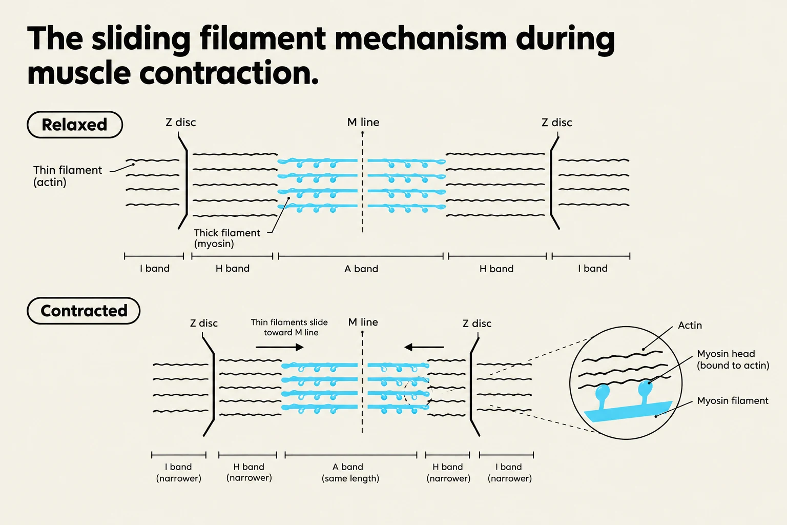
The highly organized, overlapping arrangement of these filaments creates the characteristic striations (alternating light and dark bands) seen in skeletal muscle. These bands are defined by specific landmarks:
A band (Anisotropic band): The dark band representing the entire length of the thick filaments, including the areas where they overlap with thin filaments.
H band: The lighter middle region of the A band that contains only thick filaments.
M line: A transverse protein located in the very center of the H band.
I band (Isotropic band): The light band consisting only of thin filaments.
Z disc (Z line): The protein complex that serves as the anchor for both thin and elastic filaments.
The segment of a myofibril located between two successive Z discs is called a sarcomere. The sarcomere is the functional contractile unit of the muscle fiber; when many sarcomeres shorten simultaneously, the entire muscle fiber shortens.
The Mechanism of Contraction
Diagram illustrating the sliding filament mechanism of muscle contraction, showing the relaxed and contracted states of sarcomeres with labeled components like Z discs, M line, actin, and myosin filaments.
Muscle contraction is triggered by a chemical signal. When a nerve stimulates the muscle, calcium is released into the sarcoplasm from the sarcoplasmic reticulum. This calcium binds to troponin, causing a conformational change (a change in shape). This shape change pulls tropomyosin away from the active sites on the actin filament, finally allowing the myosin heads to bind to actin.
This process is described by the sliding filament mechanism. It is a common misconception that the filaments themselves shrink or get shorter during contraction. In reality, the filaments maintain a constant length; instead, they slide past one another, increasing the degree of overlap and pulling the Z discs closer together.
Force Transfer and Structural Support
For a muscle to move a bone, the force generated within the myofibrils must be transferred to the surrounding connective tissues. This is facilitated by a protein called dystrophin. Dystrophin links the actin in the outermost myofilaments to membrane proteins that connect to the endomysium (the connective tissue surrounding the muscle fiber). This link ensures that the force of contraction is effectively transferred to the tendons and eventually the bones.
Clinical Correlation: Muscular Dystrophy
Genetic defects in the production of dystrophin lead to muscular dystrophy. Without functional dystrophin, the connection between the contractile machinery and the cell membrane is broken. During contraction, the cell membrane becomes damaged, leading to muscle cell death (necrosis) and the replacement of muscle tissue with fat and scar tissue.
Keep reading for free.
Create an account to finish this reading and make it part of your study routine.