Explores the fundamental structure of DNA, including nucleotides, the double helix, base pairing, and stability. It also covers DNA damage, hybridization, and the Sanger sequencing method.
Deoxyribonucleic acid (DNA) serves as the fundamental blueprint for life, storing the genetic information necessary for the development, functioning, and reproduction of all known living organisms. The ability of DNA to act as a stable repository of information while remaining accessible for replication and transcription is a direct consequence of its unique chemical structure and molecular geometry. To understand how DNA functions, we must look closely at its building blocks, the forces that stabilize its double-helical shape, and the methods scientists use to read its code.
The Building Blocks: Nucleotides and Bases
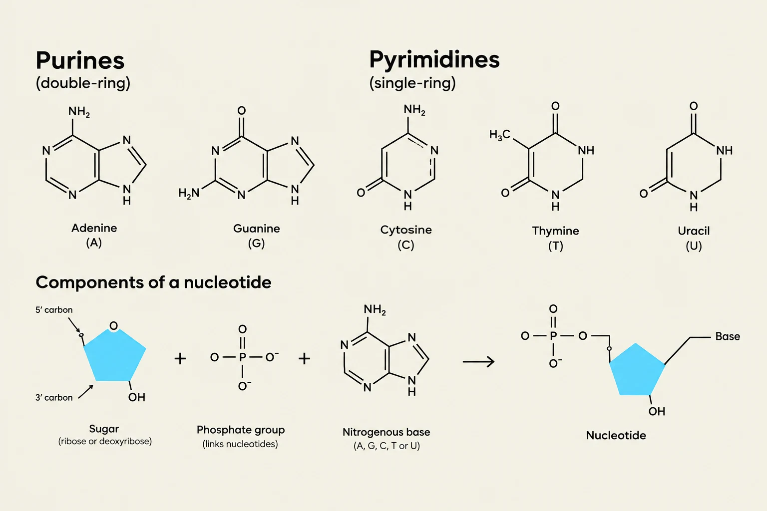
At the most basic level, DNA is a polymer made of repeating units called nucleotides. A nucleotide is a complex molecule composed of three distinct parts: a nitrogenous base, a pentose (five-carbon) sugar, and a phosphate group.
To distinguish between these components, it is helpful to understand their hierarchy:
Base: The nitrogenous ring structure that carries the genetic information.
Nucleoside: A base attached to a sugar (in DNA, this is deoxyribose) without a phosphate group.
Nucleotide: A nucleoside with an attached phosphate group.
In the context of DNA, the specific monomers used are deoxynucleoside monophosphates (dNMPs). This differs from RNA, which uses ribonucleotides (NMPs or NTPs) containing a ribose sugar.
The nitrogenous bases are categorized by their ring structures into two groups:
Purines: Double-ring structures consisting of a six-membered ring fused to a five-membered ring. These include Adenine (A) and Guanine (G).
Pyrimidines: Single-ring structures consisting of a single six-membered ring. These include Cytosine (C) and Thymine (T).
This illustration displays the chemical structures of purines (adenine and guanine) and pyrimidines (cytosine, thymine, and uracil), which are nitrogenous bases. It also shows the components of a nucleotide: a sugar (ribose or deoxyribose), a phosphate group, and a nitrogenous base, and how they combine to form a nucleotide.
The Double Helix: Stability and Polarity
DNA typically exists as a double helix (specifically the B-form at physiological pH). This structure consists of two strands that are antiparallel, meaning they run in opposite directions relative to their $5'$ and $3'$ ends.
The strands are linked by a sugar-phosphate backbone, where the phosphate group of one nucleotide is connected to the sugar of the next via a $3',5'$-phosphodiester bond. This bond formation is a critical biochemical process: during DNA synthesis, the $3'-OH$ group of the growing chain acts as a nucleophile, attacking the incoming phosphate to extend the chain.
Exam Tip: Always remember that DNA polymerase requires a free $3'-OH$ group to add the next nucleotide. Without this "hook," the chain cannot grow!
The two strands are held together by Watson-Crick base pairing. This specificity is driven by hydrogen bonding between complementary bases:
Adenine (A) pairs with Thymine (T) via two hydrogen bonds.
Guanine (G) pairs with Cytosine (C) via three hydrogen bonds.
While hydrogen bonding provides specificity, the overall stability of the helix is heavily supported by base stacking interactions. These are non-covalent forces (including hydrophobic interactions and van der Waals forces) that occur between the flat surfaces of adjacent bases stacked on top of one another along the same strand.
Thermal Properties and Secondary Structures
Because the two strands are held together by relatively weak non-covalent interactions, they can be separated. This process is called denaturation. Denaturation can be induced by high temperatures or extreme pH changes. Importantly, denaturation is a reversible process; if conditions return to physiological norms, the strands can "re-anneal" or hybridize.
A key metric in molecular biology is the melting temperature (Tm), which is the temperature at which 50% of the DNA strands are denatured. Because G-C pairs have three hydrogen bonds compared to the two in A-T pairs, DNA with a higher G-C content will have a higher Tm.
Beyond the standard helix, single strands of DNA can fold back on themselves to form various secondary structures if they contain complementary sequences. Common structures include:
Hairpins: A single loop formed by a short stem of paired bases.
Cruciforms: Formed from palindromic sequences (sequences that read the same forward and backward on complementary strands).
Mirror repeats: Sequences that are reflections of each other.
DNA Damage and Hybridization
DNA is not invincible. It is subject to spontaneous chemical changes, such as deamination (e.g., Cytosine converting to Uracil) and depurination (the loss of a purine base). While cells have sophisticated repair mechanisms, these events can lead to mutations if left unchecked.
On the positive side, the ability of two near-complementary strands to find each other and pair up—a process called DNA hybridization—is a cornerstone of biotechnology. Hybridization is used for:
Detection: Identifying specific sequences using radioactive probes or fluorescent DNA chips.
Amplification: Techniques like PCR (Polymerase Chain Reaction) rely on hybridization to prime new DNA strands.
Therapy and Evolution: Studying evolutionary relationships or developing antisense therapy.
Determining the Code: The Sanger Procedure
To make use of DNA, we must be able to read its sequence. The Sanger sequencing method (or dideoxy method) revolutionized this by using controlled premature strand termination.
The "trick" lies in the use of dideoxynucleotides (ddNTPs). Unlike standard dNTPs, ddNTPs lack the $3'-OH$ group required to form the next phosphodiester bond. When a DNA polymerase incorporates a ddNTP, the chain is terminated immediately.
Evolution of the Method
Traditional Sanger Method: This required four separate reaction tubes, one for each of the four ddNTPs. A small amount of ddNTP was added to a large pool of standard dNTPs so that termination happened randomly at different lengths. The resulting fragments were then separated by size using gel electrophoresis.
Modern Sanger Method: Today, scientists use a single reaction tube containing all four standard dNTPs and fluorescently-labeled ddNTPs. Each of the four ddNTPs (A, T, C, G) is tagged with a different colored fluorescent dye. After the reaction, the fragments are separated by size via capillary electrophoresis, and a laser detector reads the color of the terminal ddNTP to determine the exact sequence of bases.
Comparison of the traditional Sanger method, which uses four separate reactions, and the modern Sanger method, which uses a single tube for DNA sequencing.
Keep reading for free.
Create an account to finish this reading and make it part of your study routine.