Structural and Functional Classifications of Articulations
An overview of how joints are classified based on their function (degree of movement) and structure (material binding the bones), including fibrous, cartilaginous, and synovial joints, and common injuries.
In the human body, bones do not simply float independently; they are connected at specific points known as articulations, or joints. These joints serve two vital, interconnected purposes: they hold the skeletal components together to provide structure, and they allow for the mobility required for movement. To understand how the body moves and stays stable, anatomists categorize joints using two distinct systems: functional classification, which looks at how much movement is allowed, and structural classification, which looks at what material is binding the bones together.
Functional Classification: How Much Can It Move?
The functional classification of a joint is determined by the degree of movement it permits. This is a practical way of looking at joints—for example, your skull needs to be rigid for protection, while your shoulder needs to be highly mobile for reaching.
There are three primary functional categories:
Synarthroses: These are immovable joints. They provide maximum stability and protection, such as the sutures found between the bones of the skull.
Amphiarthroses: These are slightly movable joints. They offer a balance between stability and flexibility, such as the intervertebral discs in your spine or the joints between your ribs.
Diarthroses: These are freely movable joints. These are the most common joints in the limbs, including the shoulder, hips, knees, and elbows.
Exam Tip: When studying for exams, remember that "Syn-" often implies "together/fixed" (immovable), "Amphi-" implies "both/partial" (slightly movable), and "Di-" implies "through/apart" (freely movable).
Structural Classification: What Holds Them Together?
While functional classification tells us how a joint moves, structural classification tells us what the joint is made of. This classification is based on the type of binding material used and whether or not a joint cavity (a fluid-filled space) is present between the bones.
Fibrous Joints
Fibrous joints are characterized by the absence of a joint cavity. Instead, the bones are held together by dense fibrous connective tissue. The amount of movement possible in these joints depends heavily on the length of the connective tissue fibers.
There are three specific types of fibrous joints:
Sutures: These are "seams" found exclusively between the bones of the skull. Their primary job is to protect the brain tissue. As a person reaches full growth, these fibrous tissues often ossify, causing the skull bones to fuse together.
Syndesmoses: In these joints, bones are connected by a ligament. The range of movement can vary from very little to considerable depending on the length of the ligament. For example, the distal ends of the tibia and fibula are connected by a short, nearly immovable syndesmosis, whereas the long interosseous membrane between the radius and ulna allows for the rotation of the radius.
Gomphoses: This is a "peg-in-socket" joint. The most notable example is the connection between a tooth and its bony socket, held in place by the very short periodontal ligament.
The image illustrates three types of fibrous joints: sutures, syndesmosis, and gomphosis, with diagrams and brief descriptions of each.
Cartilaginous Joints
Like fibrous joints, cartilaginous joints lack a joint cavity. However, instead of fibrous tissue, the articulating bones are united by cartilage. These are divided into two types:
Synchondroses: These are immovable joints where a bar or plate of hyaline cartilage unites the bones. An example is the epiphyseal plate in long bones, which connects the diaphysis and epiphysis; these are temporary joints that ossify once adult growth is complete. Another example is the connection between the first rib and the manubrium of the sternum.
Symphysis: These are slightly movable joints. In a symphysis, the articular surfaces of the bones are covered with hyaline cartilage, which is then fused to a layer of fibrocartilage. The fibrocartilage acts as a vital shock absorber. Common examples include the intervertebral joints and the pubic symphysis.
Synovial Joints
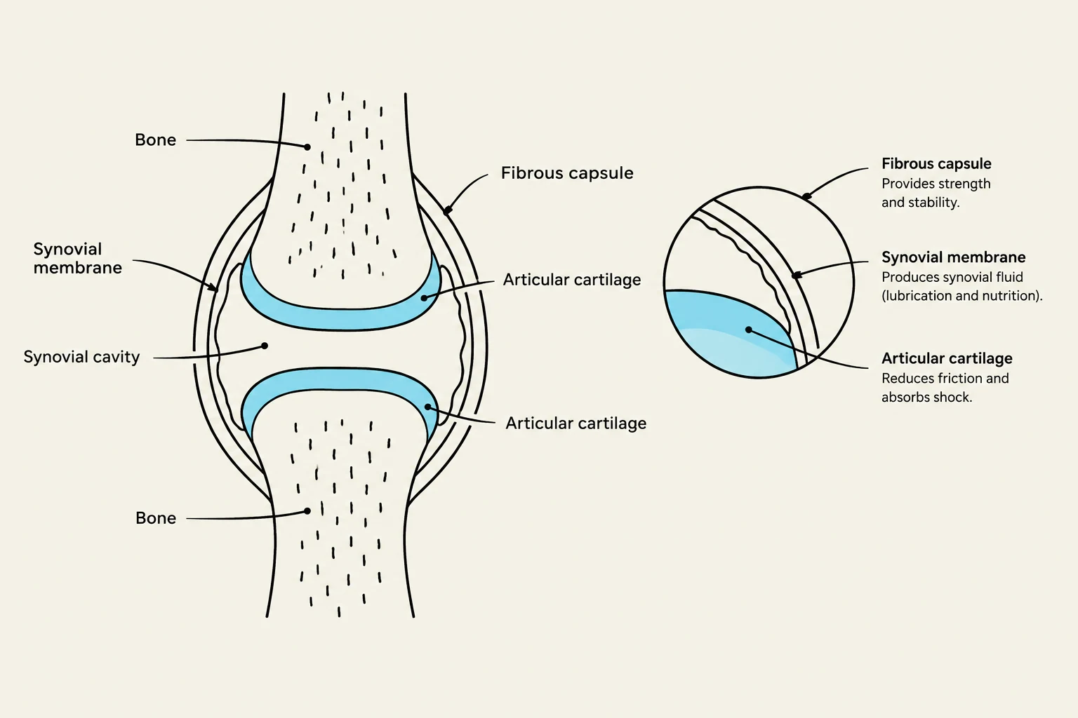
Synovial joints are the most complex and mobile structural category. Unlike fibrous and cartilaginous joints, synovial joints are distinguished by the presence of a joint (synovial) cavity—a potential space between the bones that contains a small amount of synovial fluid.
Diagram illustrating the anatomy of a synovial joint, including bones, articular cartilage, fibrous capsule, synovial membrane, and synovial cavity.
All synovial joints share five distinguishing features:
Articular Cartilage: A layer of glassy-smooth hyaline cartilage that covers the opposing bone surfaces to reduce friction.
Joint (Synovial) Cavity: The space containing synovial fluid.
Articular Capsule: A two-layered enclosure. The external layer is a fibrous capsule made of dense irregular connective tissue that strengthens the joint. The inner layer is the synovial membrane, a loose connective tissue that produces the synovial fluid.
Synovial Fluid: A slippery, viscous fluid that occupies the joint space. It reduces friction and is often described as "weeping lubrication" because it is forced from the cartilage when the joint is compressed and seeps back in when pressure is relieved.
Reinforcing Ligaments: Band-like structures that strengthen the joint.
To further assist with movement and stability, synovial joints often include bursae—flattened fibrous sacs lined with synovial membrane that act like "ball bearings" to reduce friction where muscles, tendons, or skin rub against bone.
Stability and Range of Motion
The ability of a joint to move (its Range of Motion or ROM) is determined by the structure of the articular surfaces, the strength of the ligaments and tendons, and the action of the muscles.
Stability is a balancing act. While the shape of the bones (like the deep socket of the hip) plays a role, the most important factors for stability are ligaments and muscle tone. Tendons crossing a joint, kept taut by muscle tone, provide the most significant reinforcement for joints like the shoulder and knee.
Clinical Note: If a muscle or its associated tendon does not cross a joint, that joint will not experience movement from that muscle.
Common Joint Injuries and Conditions
Because joints are essential for movement, they are susceptible to various injuries and diseases:
Sprains and Strains: A sprain is the stretching or tearing of ligaments, while a strain is the stretching or tearing of tendons.
Dislocations: This occurs when bones are forced out of alignment. A total dislocation is called a luxation, while a partial dislocation is a subluxation.
Inflammatory Conditions: Bursitis is the inflammation of a bursa (often from friction), and tendonitis is the inflammation of tendon sheaths.
Arthritis: This is a broad term for inflammatory or degenerative diseases.
Osteoarthritis (OA): A "wear-and-tear" disease where articular cartilage is destroyed faster than it can be replaced.
Rheumatoid Arthritis (RA): An autoimmune disorder where the immune system attacks its own joint tissues.
Gouty Arthritis: Caused by uric acid crystals depositing in the soft tissues of joints, causing intense pain.
When joints become too diseased to function, medical intervention such as arthroplasty—the replacement of a diseased joint with an artificial prosthesis—may be required.
Keep reading for free.
Create an account to finish this reading and make it part of your study routine.