Standardization of Immunohistochemical Staining Methods
This document explains the critical importance of standardizing immunohistochemistry (IHC) and in situ hybridization (ISH) techniques in pathology laboratories. It details how pre-analytical variables, such as fixation and tissue processing, significantly impact diagnostic accuracy. The text also covers predictive and
In the modern pathology laboratory, the ability to visualize specific proteins and nucleic acids within tissue sections is a cornerstone of precision medicine. Immunohistochemistry (IHC) and In Situ Hybridization (ISH) allow clinicians to look beyond general cell morphology to identify the molecular drivers of disease. However, the transition from a tissue biopsy to a diagnostic result is fraught with potential errors. Because these techniques are increasingly used to guide life-altering treatments, the standardization of all variables—particularly those occurring before the staining begins—is not merely a matter of laboratory efficiency; it is a fundamental requirement for patient safety.
The Clinical Stakes: Predictive and Prognostic Markers
The primary driver behind the push for rigorous standardization is the shift in how biomarkers are used in clinical practice. We no longer use IHC solely to identify the type of tumor a patient has; we use it to determine how that tumor will behave and which drugs will work.
Biomarkers are generally categorized into two functional roles:
Prognostic Markers: These provide information about the likely course of the disease (e.g., how aggressive a tumor is) regardless of the treatment chosen.
Predictive Markers: These are perhaps more critical for modern oncology. They identify whether a patient is likely to respond to a specific targeted therapy.
For example, in breast cancer diagnostics, several key proteins serve as essential predictive markers:
HER2 (human epidermal growth factor receptor 2): High expression of this protein indicates that a patient may benefit from HER2-targeted therapies, such as trastuzumab.
ER (Estrogen Receptor) and PgR (Progesterone Receptor): The presence of these receptors helps clinicians decide if hormone therapy will be an effective treatment strategy.
Clinical Note: An error in the IHC staining of HER2 can lead to a "false negative," where a patient is denied a life-saving targeted therapy, or a "false positive," where they are subjected to unnecessary toxicity from a drug that will not work.
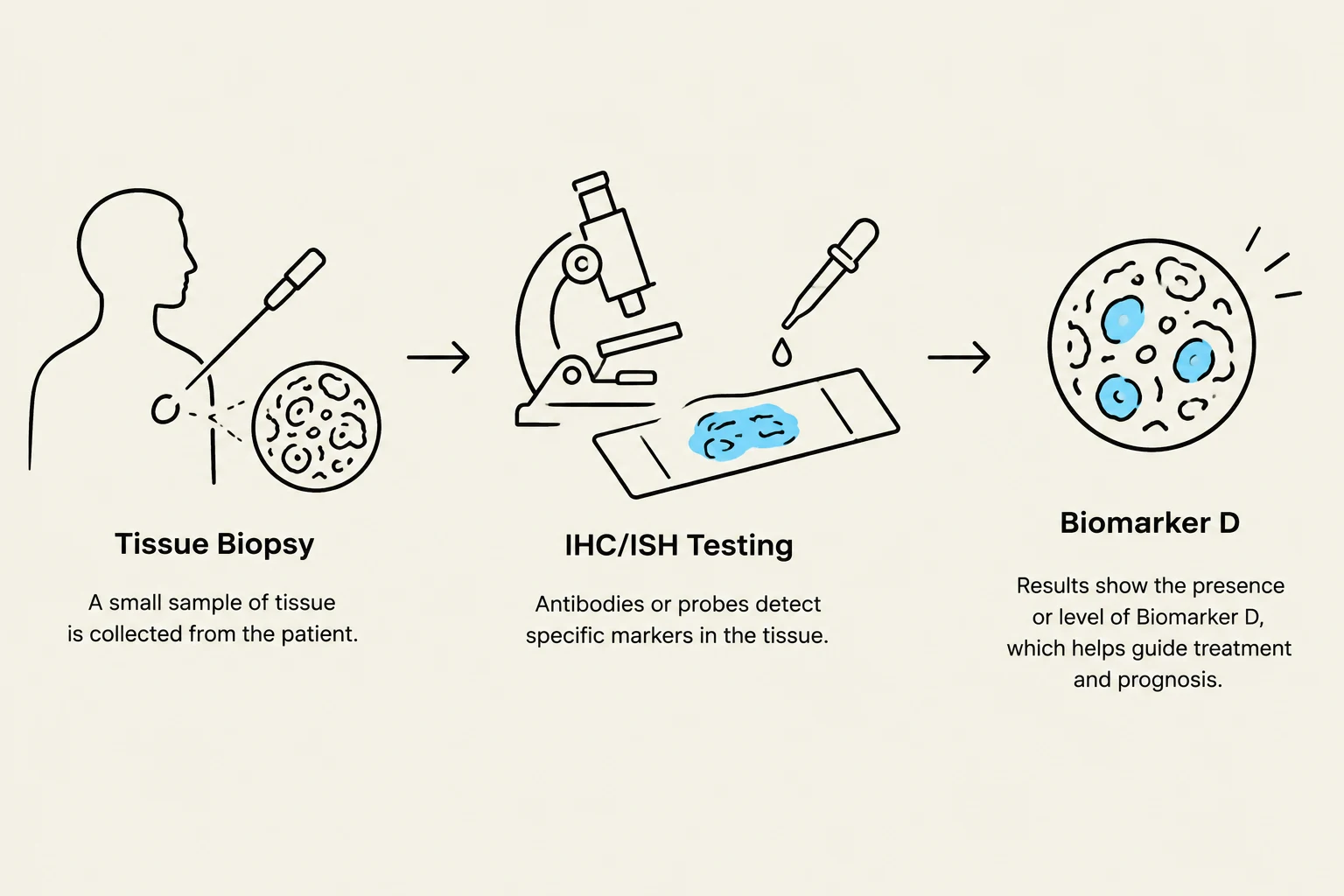
This diagram illustrates the process of tissue biopsy, IHC/ISH testing, and biomarker identification, which aids in guiding treatment and prognosis.
The Challenge of Pre-analytical Variables
The most significant threat to the accuracy of IHC and ISH results lies in the "pre-analytical" phase. These are the steps that occur before the actual staining protocol is initiated in the laboratory. If the tissue is not handled correctly at the start, even the most expensive and advanced automated stainer cannot produce a reliable result.
Fixation and Tissue Processing
Fixation is the most critical pre-analytical step. The goal of fixation, typically using formalin, is to preserve the tissue's structure and "freeze" the proteins (antigens) in place so they can be detected later.
Several factors can compromise this process:
Timing: If fixation is too short, the tissue may undergo autolysis (self-digestion), destroying the very proteins we want to see. If fixation is too long, the proteins may become overly cross-linked, making them impossible to detect.
Ischemic Time: The time elapsed between removing the tissue from the body and placing it in fixative is vital.
Tissue Processing: The method used to create Formalin-Fixed, Paraffin-Embedded (FFPE) blocks must be consistent to prevent artifacts or the loss of protein expression.
Antigen Retrieval (AR)
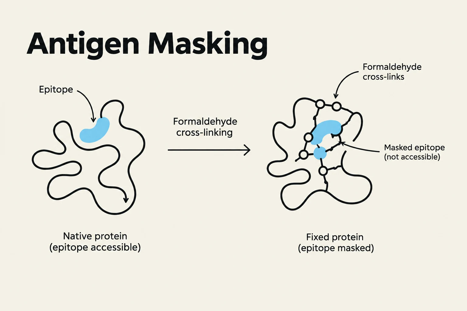
During the fixation process, the chemical bonds formed can "mask" the epitopes (the specific parts of the protein that antibodies recognize). To fix this, we use Antigen Retrieval to uncover these hidden sites. This is usually achieved through:
Heat-Induced Epitope Retrieval (HIER): Using heat (via microwave, autoclave, or pressure cooker) in a specific buffer solution.
Proteolytic Digestion: Using enzymes (like pepsin) to digest the protein mask around the epitope.
This diagram illustrates antigen masking, where formaldehyde cross-linking modifies a protein, making its epitope inaccessible.
Achieving Methodological Consistency
While it is technically impossible to eliminate every single variable in a biological system, laboratories achieve reliability through standardization.
Standardization does not mean that every lab must use the exact same machine, but rather that every lab must follow a rigorous, validated protocol. This involves:
Protocol Optimization: Determining the exact concentration of antibodies, the precise pH of buffers, and the specific timing for each step.
Validation and Verification: Proving that a protocol works as intended and produces reproducible results across different batches of tissue.
Use of Controls: Running "control" tissues (known positive and negative samples) alongside patient samples to ensure the staining worked correctly.
When a laboratory adheres to a strict protocol, it creates a "baseline." If a result looks strange, the pathologist can look back at the standardized steps to troubleshoot whether the issue was the antibody, the fixation, or the retrieval process.
Summary of the Standardization Workflow
To visualize the complexity, consider the following relationship between variables and the final diagnostic output:
By minimizing the denominator (variability) through strict adherence to fixation and processing guidelines, the laboratory maximizes the reliability of the clinical result.
Exam Tip: If asked why standardization is critical for IHC, do not just say "for better results." The most complete answer is: "To ensure the reliable measurement of predictive biomarkers (like HER2, ER, and PgR), which directly informs clinical decision-making and patient safety".
Keep reading for free.
Create an account to finish this reading and make it part of your study routine.