


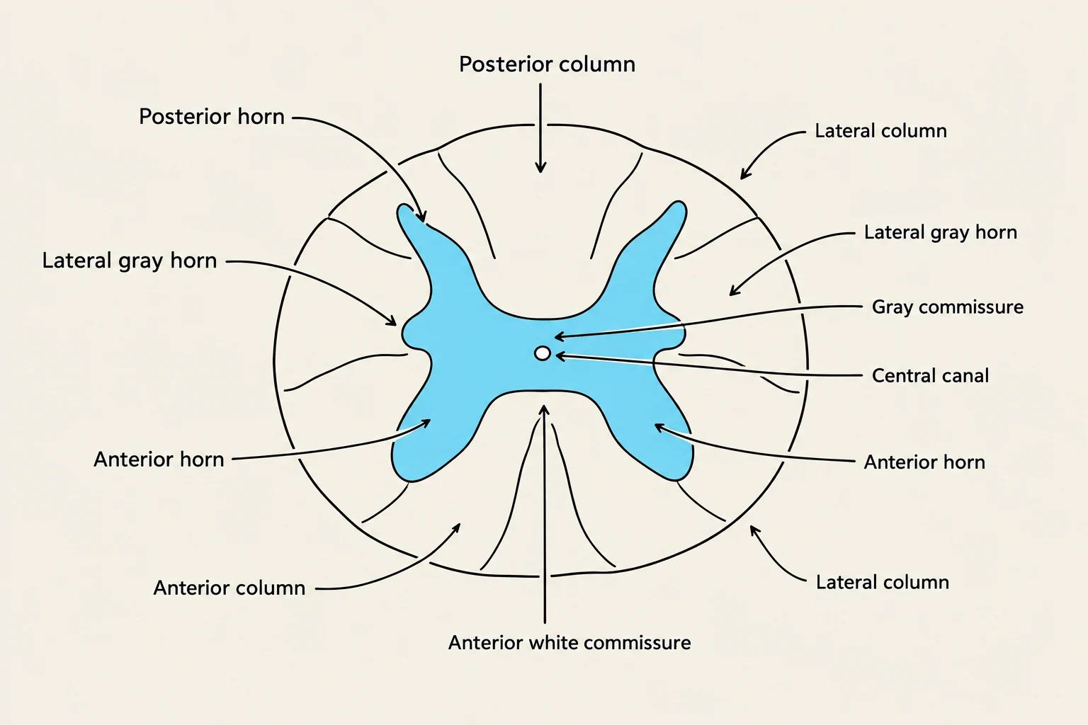
The spinal cord serves as the vital communication highway between the brain and the rest of the body. It is not merely a passive cable; it is a complex structure of specialized tissues that process sensory information and coordinate motor responses. To understand how the spinal cord functions, one must look at its protective layers, its external nerve connections, and its intricate internal organization of gray and white matter.

The Protective Layers: The Meninges
The spinal cord is highly delicate and requires multiple layers of protection to shield it from physical and chemical injury. These layers, known as the meninges, are continuous with the meninges that surround the brain.
The Three Layers
From the outermost layer to the innermost, the meninges consist of:
- Dura mater: This is the most superficial and toughest layer. In the spinal cord, the dura mater forms a protective sac that extends from the foramen magnum down to the second sacral vertebra.
- Arachnoid mater: The middle layer, which is thinner than the dura.
- Pia mater: The innermost layer. It is a transparent membrane that adheres directly to the surface of the spinal cord, carrying blood vessels that provide essential nutrients to the neural tissue.
The Fluid-Filled Spaces
Between these membranes lie specific spaces that serve critical physiological roles:
- Epidural space: Located between the dura mater and the vertebral canal, this space contains fat and connective tissue that provide cushioning for the cord.
- Subdural space: A thin space containing interstitial fluid that lies between the dura mater and the arachnoid mater.
- Subarachnoid space: This space lies between the arachnoid mater and the pia mater. It is filled with cerebrospinal fluid (CSF).

Keep reading for free.
Create an account to finish this reading and make it part of your study routine.
Continue reading freeAlready have an account? Log in
Updated