This content explains the anatomy of the spinal cord, including its structure, spinal nerves, and innervation patterns. It also details the primary and secondary mechanisms of spinal cord injury, common clinical syndromes resulting from such injuries, and acute complications like spinal shock and autonomic hyperreflexi
The spinal cord serves as the vital communication highway of the central nervous system, facilitating the bidirectional flow of nervous impulses between the brain and the rest of the body. Beyond simple transmission, it acts as a sophisticated processing center for spinal reflexes, managing essential functions such as posture maintenance, protective responses to pain, muscle tone, and urination. Understanding its intricate anatomy and the complex mechanisms of injury is fundamental to clinical neurology and emergency medicine.
Structural Anatomy of the Spinal Cord
The spinal cord is housed within the vertebral column, a protective structure formed by interlocking bones. These bones are cushioned by intervertebral disks to absorb shock and provide flexibility. For the nervous system to communicate with the periphery, the spinal nerves must exit this bony protection through specific openings known as the intervertebral foramina.
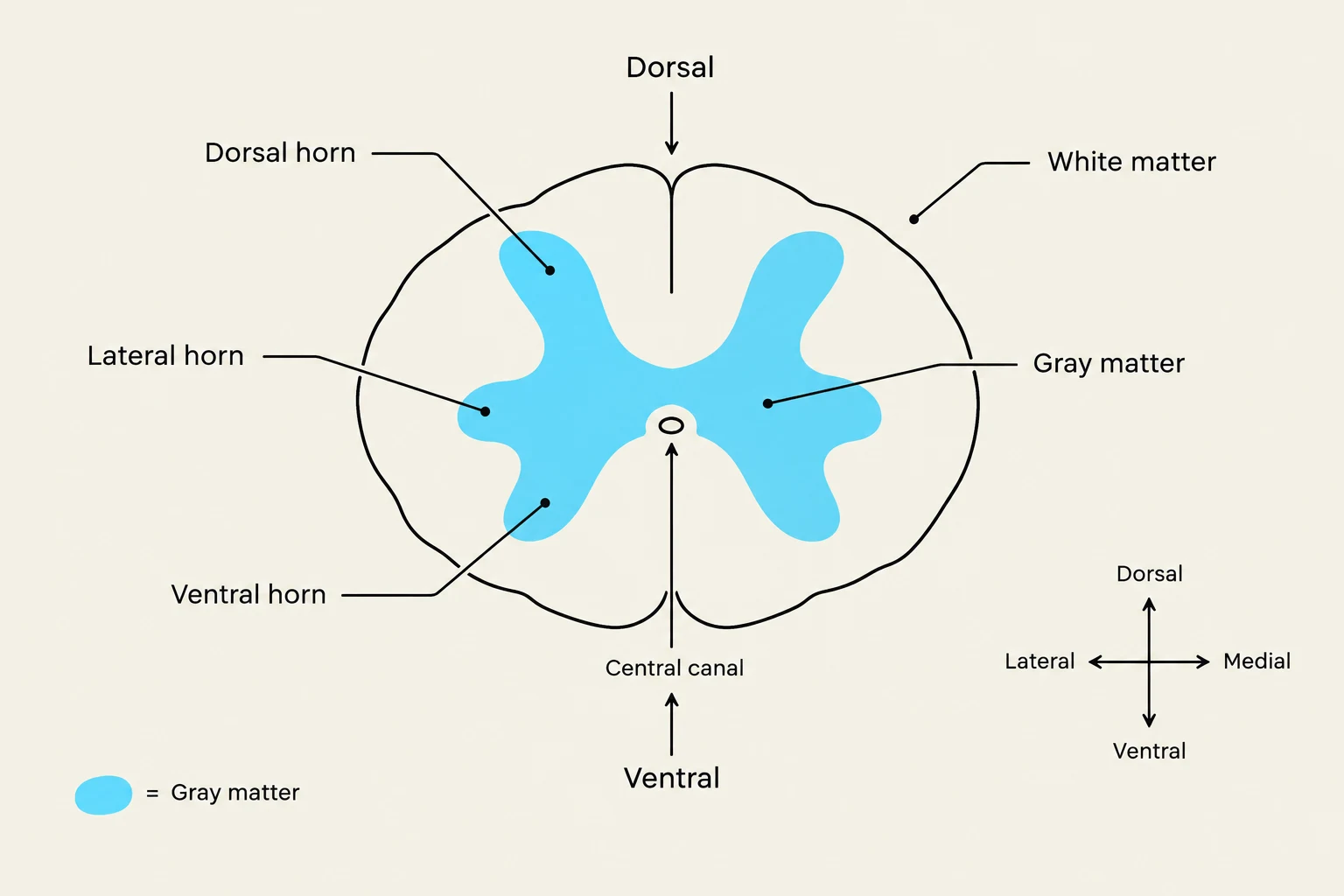
Internally, the spinal cord exhibits a distinct organization of matter. It features a "butterfly" pattern of gray matter centrally located, surrounded by an outer layer of white matter. This gray matter is organized into specific functional "horns":
Dorsal Horn: Contains sensory neurons that receive incoming information from the body.
Ventral Horn: Contains motor neurons that send outgoing signals to muscles.
Lateral Horn: Contains sympathetic neurons, which are part of the autonomic nervous system.
Diagram illustrating a cross-section of the spinal cord, labeling the dorsal and ventral horns, gray and white matter, and the central canal.
Spinal Nerves and Innervation Patterns
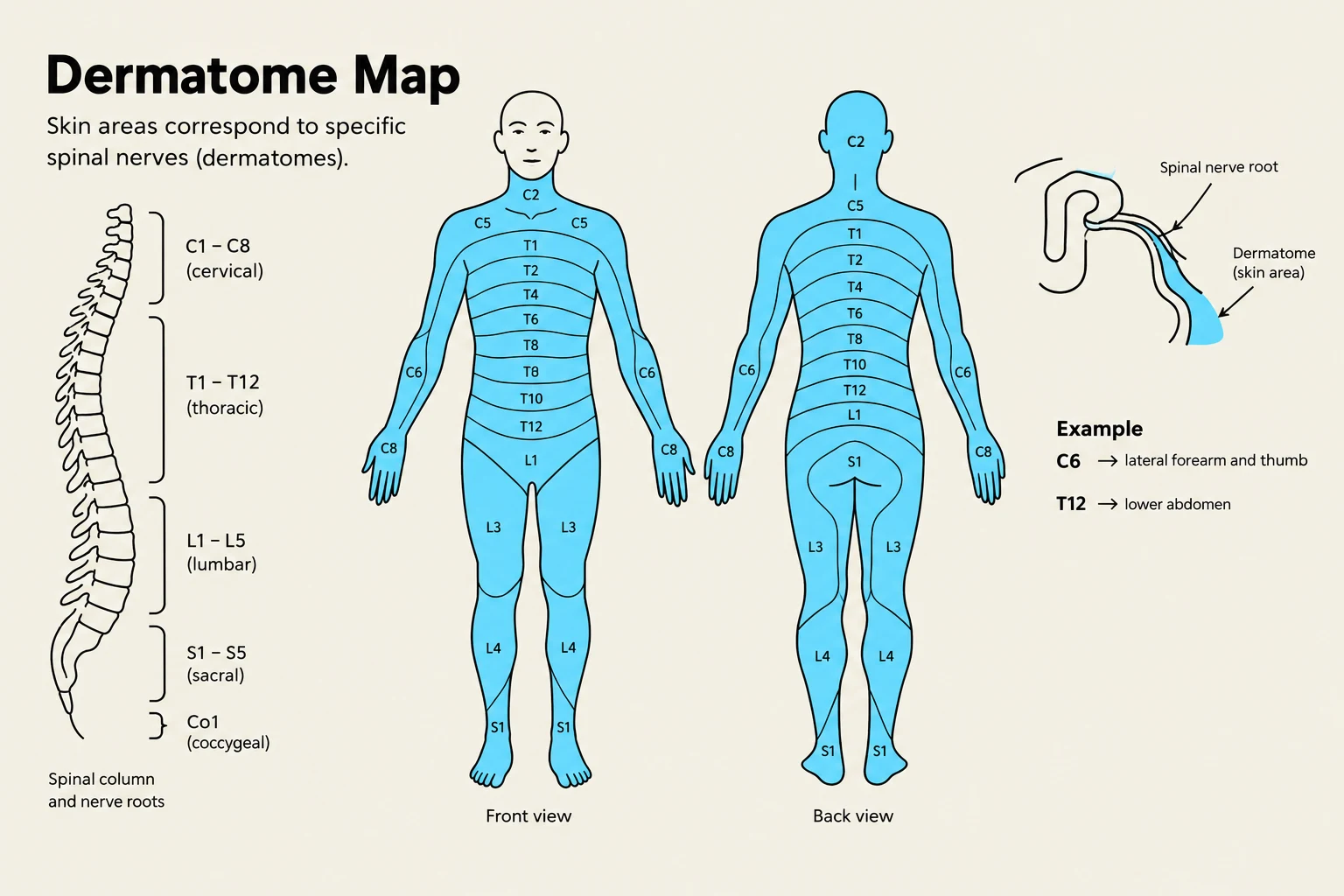
The spinal cord communicates with the body through 31 pairs of spinal nerves. These are categorized by their location: 8 cervical, 12 thoracic, 5 lumbar, 5 sacral, and 1 coccygeal pair.
These nerves carry both somatic (voluntary) and autonomic (involuntary) information. A key anatomical concept is the dermatome, which refers to the specific segment of the body's skin that is innervated by a single spinal nerve.
Most spinal nerves do not travel alone; instead, they merge to form complex networks called spinal plexuses. For example, the brachial plexus is a major network of nerves. However, there is a notable exception: the thoracic nerves from T2 to T12 do not form plexuses. Instead, they travel parallel to the ribs to innervate the intercostal muscles and the skin of the trunk and abdomen.
This diagram illustrates the dermatome map, showing how specific areas of the skin correspond to different spinal nerves. It includes front and back views of the human body, a representation of the spinal column, and an example of how dermatomes relate to body parts.
Mechanisms of Spinal Cord Injury
Spinal cord injuries (SCI) are categorized by the timing and nature of the damage. Clinicians distinguish between the immediate physical trauma and the subsequent biochemical cascade.
Primary Injury Mechanisms
Primary injury refers to the immediate mechanical damage occurring at the moment of impact. Common mechanisms include:
Shearing forces
Laceration (tearing of tissue)
Acute stretching
Sudden acceleration-deceleration forces
Secondary Injury Mechanisms
Secondary injury refers to the physiological processes that begin after the initial trauma and can exacerbate the damage over time. These include:
Biochemical/Cellular: Free radicals, apoptosis (programmed cell death), and the release of cytokines by invading immune cells.
Metabolic: Energy failure, decreased ATP production, and the desynchronization of ionic homeostasis.
Inflammatory: General inflammation and immune cell invasion.
Exam Tip: Remember that while primary injury is mechanical and instantaneous, secondary injury is a progressive biochemical cascade that can lead to further tissue loss even after the patient is stabilized.
Clinical Syndromes and Complications
Damage to specific parts of the spinal cord results in predictable patterns of neurological deficit.
Syndrome
Clinical Presentation
Central Cord Syndrome
Bilateral loss of motor function (upper extremities > lower extremities); pain/temperature sensation reduced; proprioception/vibration spared
Anterior Cord Syndrome
Loss of motor function, touch, pain, and temperature below the lesion; proprioception and vibration remain intact
Brown-Séquard Syndrome
Ipsilateral (same side) loss of motor, touch, proprioception, and vibration; contralateral (opposite side) loss of pain and temperature
Case Study: Brown-Séquard Syndrome
A 25-year-old woman suffers a puncture wound to the neck. A CT scan reveals the injury pierced the first thoracic vertebra. She presents with right spastic hemiparaplegia and asymmetrical sensory loss: she has lost vibration and proprioception on the right side, but has lost pain and temperature sensation on the left side.
Diagnosis: This is a classic presentation of Brown-Séquard syndrome, caused by a right hemisection (half-cut) of the spinal cord.
Acute Complications
Following an injury, patients may experience several life-threatening complications:
Spinal Shock: A temporary loss of all reflexes and sensorimotor function below the level of injury.
Neurogenic Shock: Hemodynamic instability (bradycardia, hypotension, and hypoventilation) caused by the loss of sympathetic tone, typically seen in rostral (high) cord injuries.
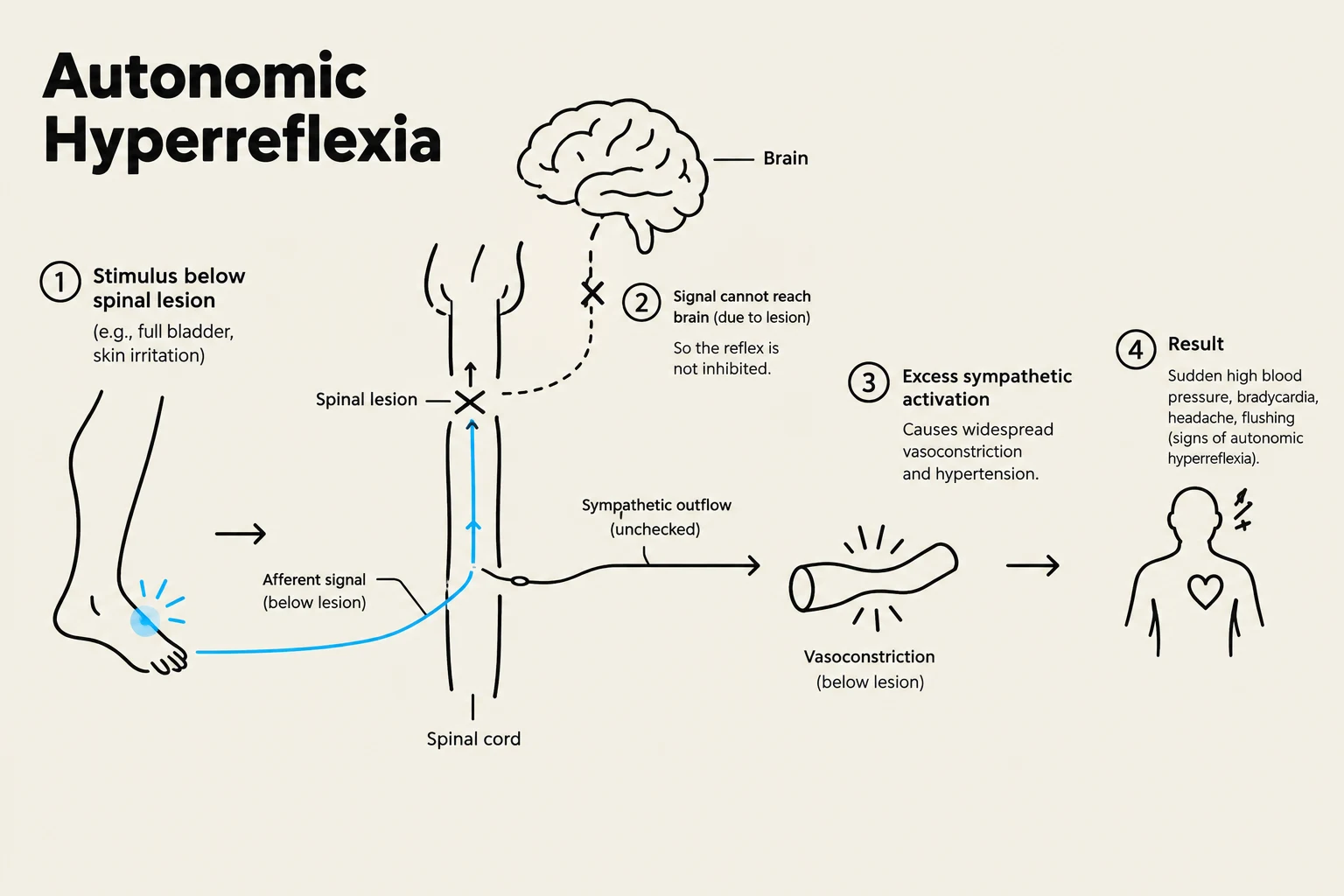
Autonomic Hyperreflexia: A potentially fatal condition in injuries at or above the T6 level. It is characterized by extreme hypertension and bradycardia due to a massive sympathetic response below the lesion and a compensatory parasympathetic response above it. Common triggers include bladder distention or UTIs.
Diagram illustrating the physiological process of autonomic hyperreflexia, a condition that can occur after spinal cord injury.
Keep reading for free.
Create an account to finish this reading and make it part of your study routine.