Small Ruminant and Camelid Clinical Procedures and Management
A comprehensive guide to the management and clinical care of small ruminants (sheep and goats) and camelids, covering breed classifications, terminology, gastrointestinal anatomy, clinical procedures like venipuncture and injections, anesthesia, pain management, vaccination protocols, parasite control, and disbudding.
Small ruminants, primarily sheep and goats, are essential components of global agriculture, valued for their meat, milk, and fiber production. For veterinary technicians and clinicians, mastering the nuances of these species requires an understanding of their specific terminology, unique gastrointestinal anatomy, and specialized clinical procedures ranging from vaccinations to epidurals. This article serves as a comprehensive guide to the management and clinical care of sheep, goats, and camelids.
Breed Classifications and Terminology
Sheep and goats are categorized based on their primary production purpose: dairy, fiber, or meat.
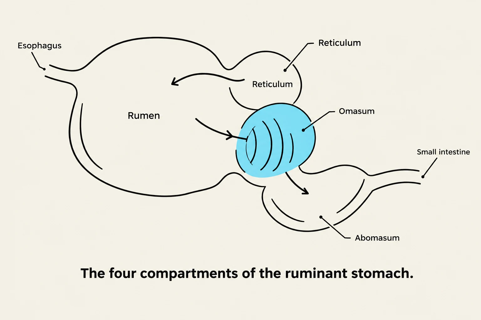
Diagram illustrating the four compartments of a ruminant stomach: esophagus, rumen, reticulum, omasum, and abomasum, leading to the small intestine.
Ruminants possess a complex four-compartment stomach designed to ferment plant material. The flow of digesta follows this path: Reticulum → Rumen → Omasum → Abomasum.
Rumen: The largest chamber. It contains "bugs" (microbes) that break down plant material into volatile fatty acids for energy.
Reticulum: Works in conjunction with the rumen; it is a key site in the digestive process.
Omasum: Often called "book pages," this compartment increases the absorption of nutrients and water.
Abomasum: Known as the "true stomach," it utilizes Hydrochloric Acid (HCl) to break down proteins before the contents move to the small intestine.
In young ruminants, the anatomy is functionally different. They possess an esophageal groove that helps direct milk to the abomasum. The distribution of milk flow changes with age:
At birth: 30% to the Reticulum, 60% to the Abomasum.
At 4 weeks: 50% to the Reticulum, 36% to the Abomasum.
At 16 weeks: 66% to the Reticulum, 15% to the Abomasum.
Clinical Note: In young ruminants, the combination of Renin and Casein leads to the formation of curd.
Clinical Procedures: Injections and Venipuncture
Venipuncture
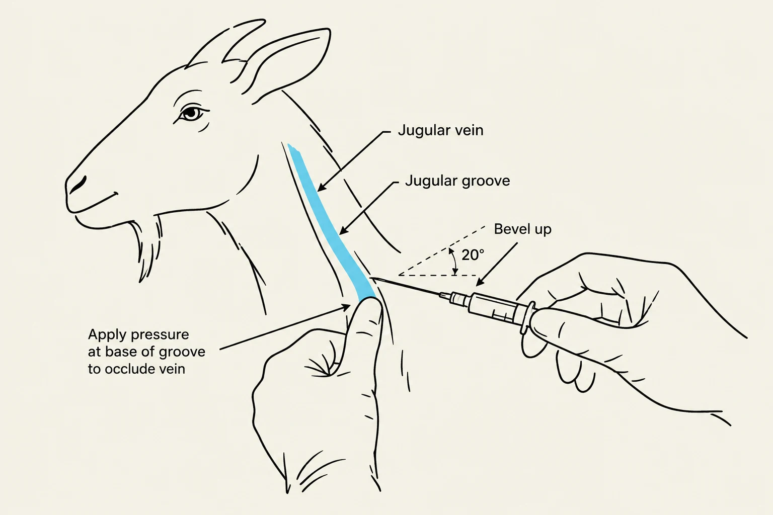
Diagram illustrating the jugular vein and groove in a goat, with instructions on applying pressure and needle insertion angle for venipuncture.
The jugular vein is the preferred site for venipuncture. To perform this:
Occlude the vein by applying pressure at the base of the jugular groove to visualize the raised vein.
Insert an 18-gauge needle at a 20° angle with the bevel facing up.
You should feel a "pop" as the needle penetrates the skin.
Pro-Tip: Unlike many other species, there is no need to clip the hair or use alcohol for routine jugular venipuncture in small ruminants.
Injections
Injections can be administered Subcutaneously (SQ) or Intramuscularly (IM). Common sites include the neck or the axillary area (the left side is typically used for goats). Needle gauge (18, 20, or 22) and length (43", 1", or 1.5") should be selected based on the specific drug and the animal's purpose.
Anesthesia and Pain Management
Sedation Pharmacology
Different drugs are selected based on the required level of sedation and the procedure being performed:
Acepromazine: Provides mild sedation; often combined with an opioid to increase depth.
Benzodiazepines (Midazolam/Diazepam): Used for mild sedation during non-painful procedures like ultrasound or radiographs. They can be reversed in newborns using Flumazenil.
Alpha-2 Adrenoceptor Agonists (Xylazine): Highly reliable but very sensitive; sheep are particularly susceptible to pulmonary edema when using this class. It can be reversed with Atipamezole.
Epidurals and Nerve Blocks
Epidurals are used to provide analgesia (pain relief) or anesthesia (loss of sensation) to specific regions.
1. Caudal Epidural
Target: Perineum, vulva, vagina, and rectum.
Uses: Castrations, prolapses, dystocia, C-sections, and vasectomies.
Procedure: Locate the 5th sacral space (where the tail articulates with the sacrum). Insert an 18-20g x 1.5-2in needle perpendicular to the spine at a 90° angle. Advance until a "pop" is felt.
Dosage: 0.5mg/kg of 2% lidocaine (without bicarbonate or epinephrine).
2. Lumbosacral Epidural
Target: Useful in sheep with ultra-short tails; provides analgesia from the caudal paralumbar space.
Uses: Dystocia, Listeria, or Meningeal Worm.
Procedure: Palpate the midline halfway between the 7th lumbar vertebra and the sacrum. Insert an 18-20g x 1.5-2in needle at 90° until a "pop" is felt.
Dosage: 0.3-0.5ml of 2% lidocaine per 10kg.
3. Local Nerve Blocks
Used during disbudding to block the cornual nerve branch of the zygomaticotemporal nerve (lateral) or the cornual branch of the infratrochlear nerve (medial).
Vaccination and Disease Management
CD&T Vaccination Protocol
The CD&T vaccine protects against Clostridium perfringens types C and D, as well as tetanus.
Does: Vaccinate 30 days prior to giving birth. Note that if the doe has not received a priming booster (two shots 3-4 weeks apart) previously, the annual shot may not be effective.
Kids: If the dam is immunized, vaccinate kids at 6 weeks and repeat at 10 weeks. If the dam is not immunized, vaccinate kids at 4, 8, and 12 weeks.
Breeding Bucks/Yearlings: Immunize at the same time as pregnant does.
Parasite Control
Haemonchus contortus (Barber Pole Worm): A blood-sucking parasite. Use the FAMACHA system to monitor for anemia and slow drug resistance.
Diagnostics: Fecal Egg Counts (FEC) using the Modified McMaster Test.
Special Considerations: Camelids
Camelids (Llamas and Alpacas) are pseudoruminants. They possess only three stomach compartments and lack the compartment that mimics the omasum.
Llama Sedation ("Llama Lullaby"): A combination of Ketamine (10ml bottle, 100mg/ml), Xylazine (1ml, 100mg/ml), and Butorphanol (1ml, 10mg/ml), dosed at 1ml per 50lbs.
Common Diseases: They are susceptible to Eimeria macusaniensis (coccidia) and Parelaphostrongylus tenuis (Meningeal Worm), which causes neurological signs.
Clinical Procedures: Disbudding
Disbudding (removing the horn bud) is preferred over dehorning.
Timing: Females (days 4-6); Males (days 3-5).
Procedure: Use hot iron cautery. Apply the circular end of the iron firmly over the bud for 10 seconds until a "copper ring" appears.
Pain Management: Use local blocks (Lidocaine 4-8 mg/kg) and systemic analgesics like Meloxicam (1mg/kg).
Keep reading for free.
Create an account to finish this reading and make it part of your study routine.