


The human body is supported by a complex framework of bones, cartilage, and connective tissues known as the skeletal system. The scientific study of bones and their associated disorders is called osteology. Far from being static, dead structures, bones are living organs that work in harmony with blood, nervous, and adipose tissues to maintain the body's integrity and internal balance.
Functions of the Skeletal System
The skeletal system serves several critical roles that are essential for survival and daily activity:
- Support: It provides the structural framework for the body.
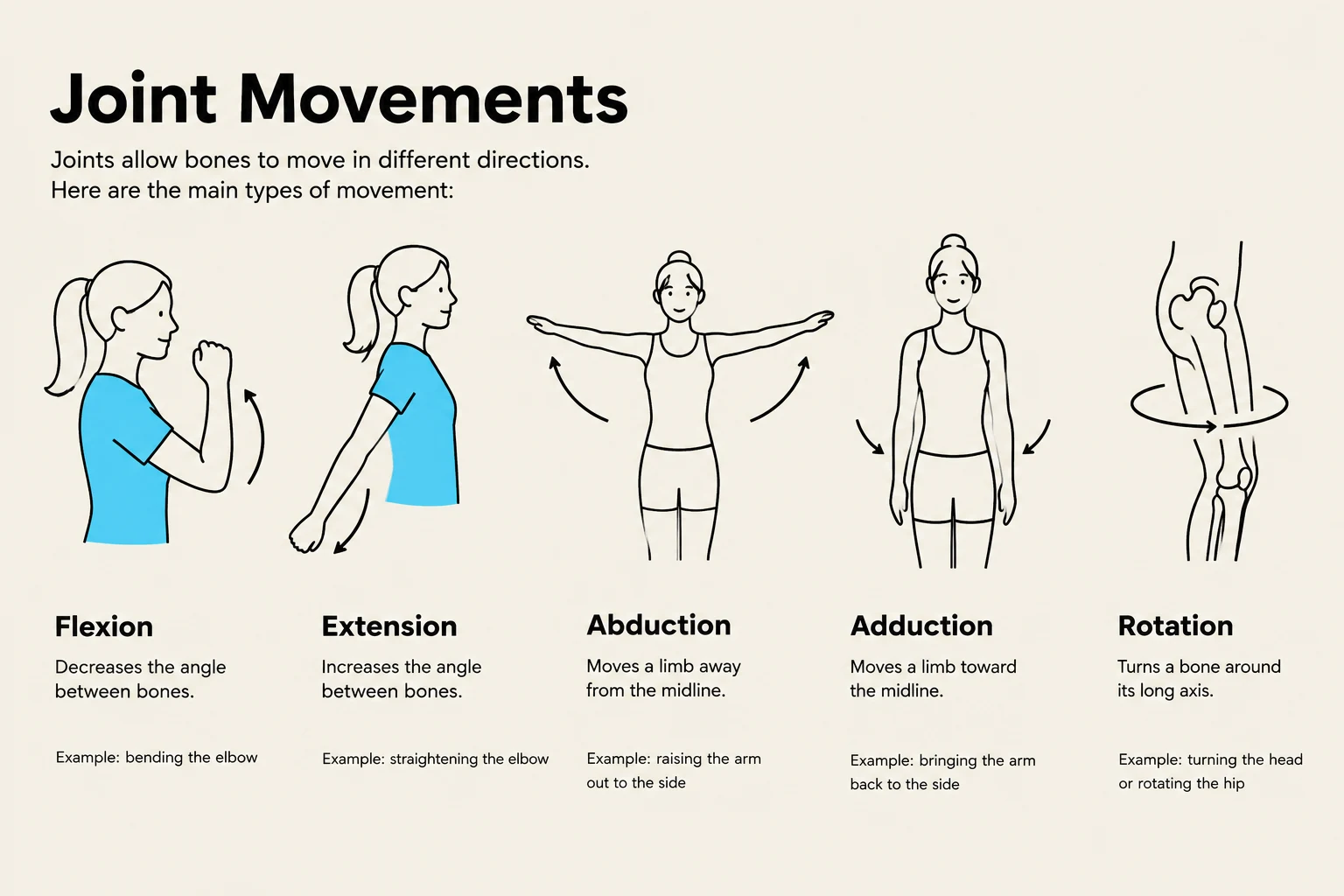
- Movement: It acts as a lever system that works with muscles to facilitate motion.
- Mineral Homeostasis: It serves as a reservoir for essential minerals, particularly calcium.
- Hemopoiesis: The process of blood cell production occurs within the bone marrow.
- Triglyceride Storage: Yellow bone marrow serves as a site for fat storage.
Anatomy of a Long Bone
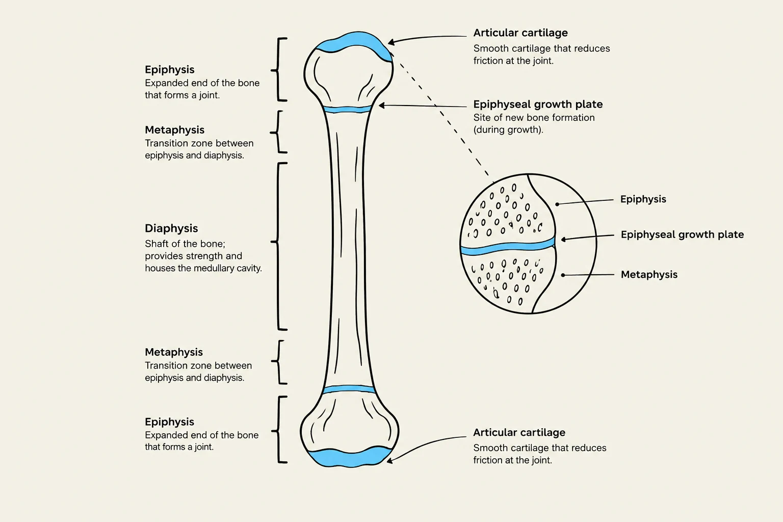
To understand how bones function, we must look at the macroscopic structure of a typical long bone (such as the femur or humerus). A long bone is composed of several distinct regions:
- Epiphyses: These are the proximal and distal ends of the bone.
- Diaphysis: This is the central shaft or the main body of the bone.
- Metaphyses: These are the transition zones located between the epiphyses and the diaphysis. Within the metaphyses lies the epiphyseal growth plate, a layer of hyaline cartilage that allows for longitudinal growth. In most individuals, this plate is replaced by bone around age 24, leaving behind an epiphyseal line.
- Articular Cartilage: A thin layer of hyaline cartilage covers the epiphyses to reduce friction during movement. Because it is avascular (lacks blood vessels) and aneural (lacks nerves), its ability to repair itself is quite limited.
- Periosteum: This is the tough, outermost connective tissue covering of the bone.
- Medullary Cavity: Located within the diaphysis, this hollow marrow cavity contains both red and yellow marrow. This cavity is essential for reducing the overall weight of the bone.
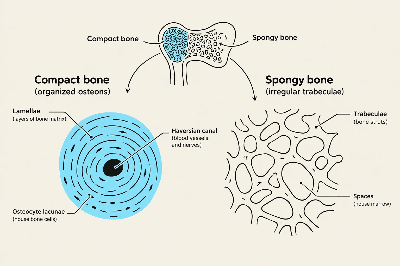
- Endosteum: A thin connective tissue membrane that lines the internal medullary cavity and the spaces within spongy bone.
Keep reading for free.
Create an account to finish this reading and make it part of your study routine.
Continue reading freeAlready have an account? Log in
Updated