Skeletal Muscle Structure, Physiology, and Contraction
An in-depth explanation of skeletal muscle structure, from the cellular level to the functional unit of the sarcomere. It details the neuromuscular junction, action potential propagation, and the process of excitation-contraction coupling.
Skeletal muscle is a highly specialized tissue designed for movement, posture maintenance, joint stabilization, and heat generation. To achieve these functions, muscle cells must convert electrical signals from the nervous system into mechanical force. This process involves a complex interplay between the nervous system and the muscle fiber's internal architecture, spanning from the microscopic synapse to the molecular level of contractile proteins.
Skeletal Muscle Structure
A single skeletal muscle cell, often called a muscle fiber, myocyte, or myofiber, is a massive cylindrical structure that can reach up to 30 cm in length. Unlike many other cells, these fibers are multinucleated and contain specialized organelles to support high metabolic demands.
The plasma membrane of the muscle fiber is known as the sarcolemma. Invaginating deep into the cell are the T-tubules (transverse tubules), which are tubular extensions of the sarcolemma that run at 90° to the fiber. These tubules are critical because they allow electrical signals to penetrate deep into the interior of the cell.
Inside the cell, the cytoplasm is called the sarcoplasm. It is packed with:
Glycosomes: Granules of stored glycogen for energy.
Myoglobin: A protein used for oxygen storage.
Myofibrils: Hundreds to thousands of rod-like contractile elements that make up about 80% of the cell volume.
Sarcoplasmic Reticulum (SR): A specialized smooth endoplasmic reticulum that surrounds each myofibril and functions to regulate intracellular Ca2+ levels.
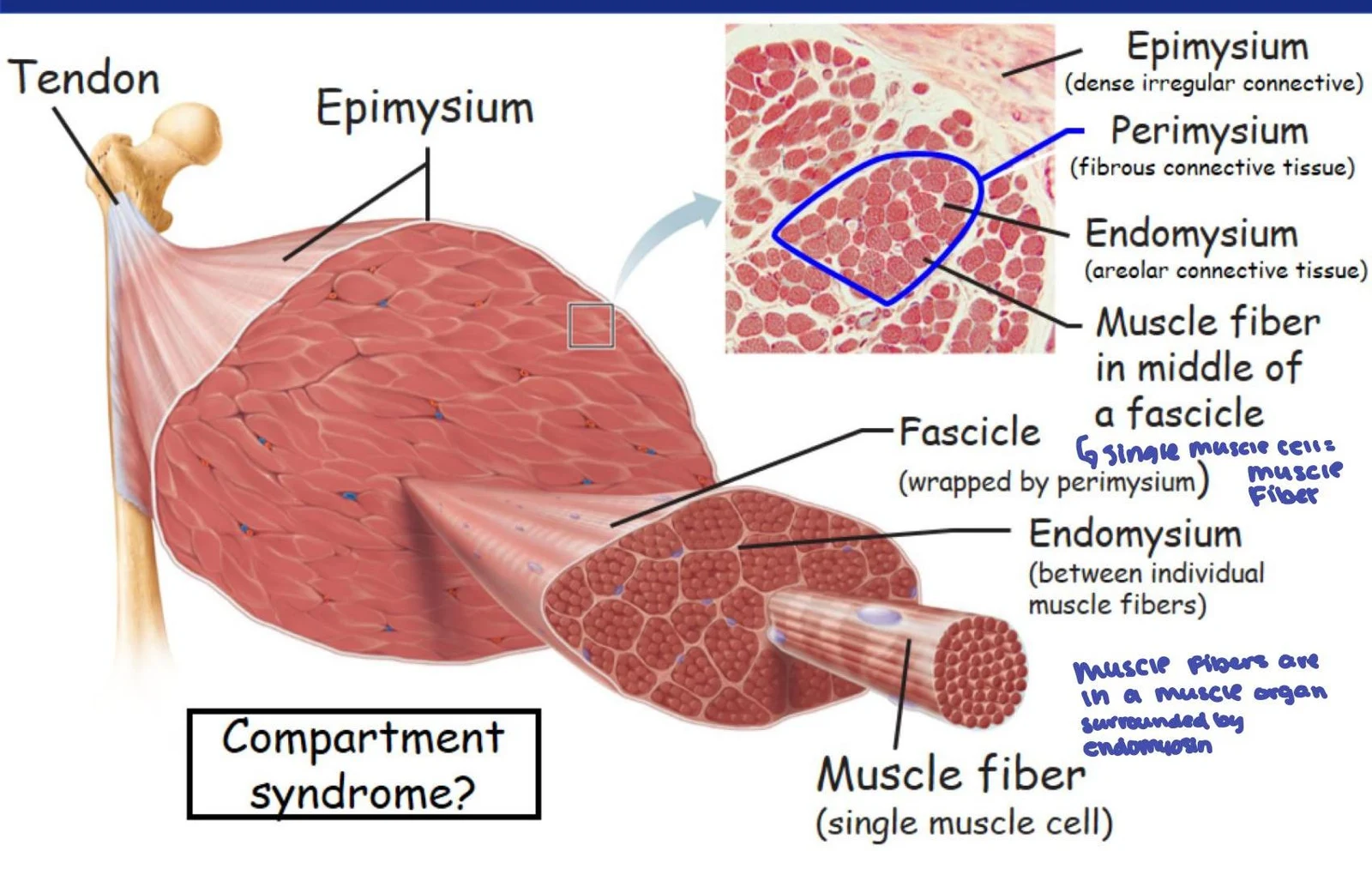
This illustration shows the structure of a muscle, including the tendon, epimysium, perimysium, endomysium, fascicle, and muscle fiber. It also includes a microscopic view of muscle tissue and labels for different connective tissues.
The Sarcomere: The Functional Unit
The myofibrils are composed of repeating units called sarcomeres, which are the smallest functional units of contraction. A sarcomere is defined as the region between two successive Z discs.
Diagram illustrating the structure of a sarcomere, the basic contractile unit of muscle tissue, showing the arrangement of actin and myosin filaments, Z discs, H zone, I band, A band, and M line.
The sarcomere contains two main types of myofilaments:
Thick Filaments (Myosin): These contain motor proteins with "heads" that possess binding sites for actin and ATP, as well as ATPase enzymes.
Thin Filaments (Actin): These consist of globular (G) actin subunits and are associated with regulatory proteins, tropomyosin and troponin.
The Neuromuscular Junction (NMJ)
Muscle activation begins at the neuromuscular junction, a specialized synapse located midway along the length of a muscle fiber where a motor neuron meets the muscle cell.
The process follows a specific sequence:
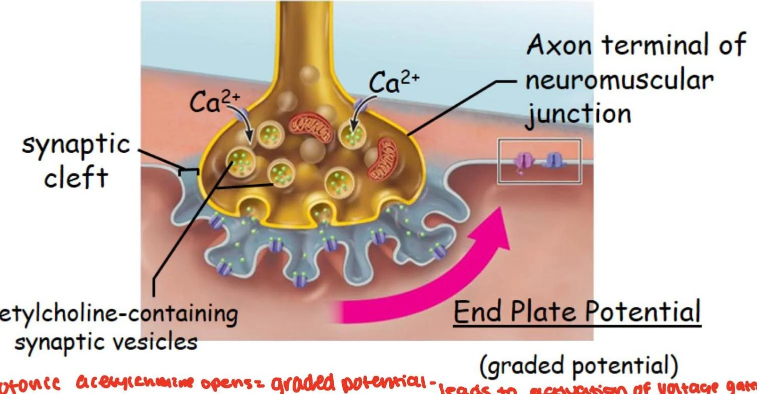
Release of Acetylcholine (ACh): When a nerve impulse reaches the axon terminal, it triggers the release of the neurotransmitter acetylcholine from synaptic vesicles into the synaptic cleft (the space between the neuron and the muscle).
Receptor Binding: ACh diffuses across the cleft and binds to nicotinic acetylcholine receptors (nAChR) located on the sarcolemma. These are ionotropic, ligand-gated channels.
End Plate Potential (EPP): The binding of two ACh molecules to the nAChR opens the channel, allowing a flow of ions (primarily Na+ influx) that creates a local, graded depolarization known as the End Plate Potential.
Diagram illustrating the neuromuscular junction, showing the release of acetylcholine from synaptic vesicles into the synaptic cleft, leading to the End Plate Potential.
Note on Regulation: To prevent continuous contraction, acetylcholine is rapidly removed from the synapse by the enzyme acetylcholinesterase (also known as cholinesterase).
Muscle Action Potentials and Propagation
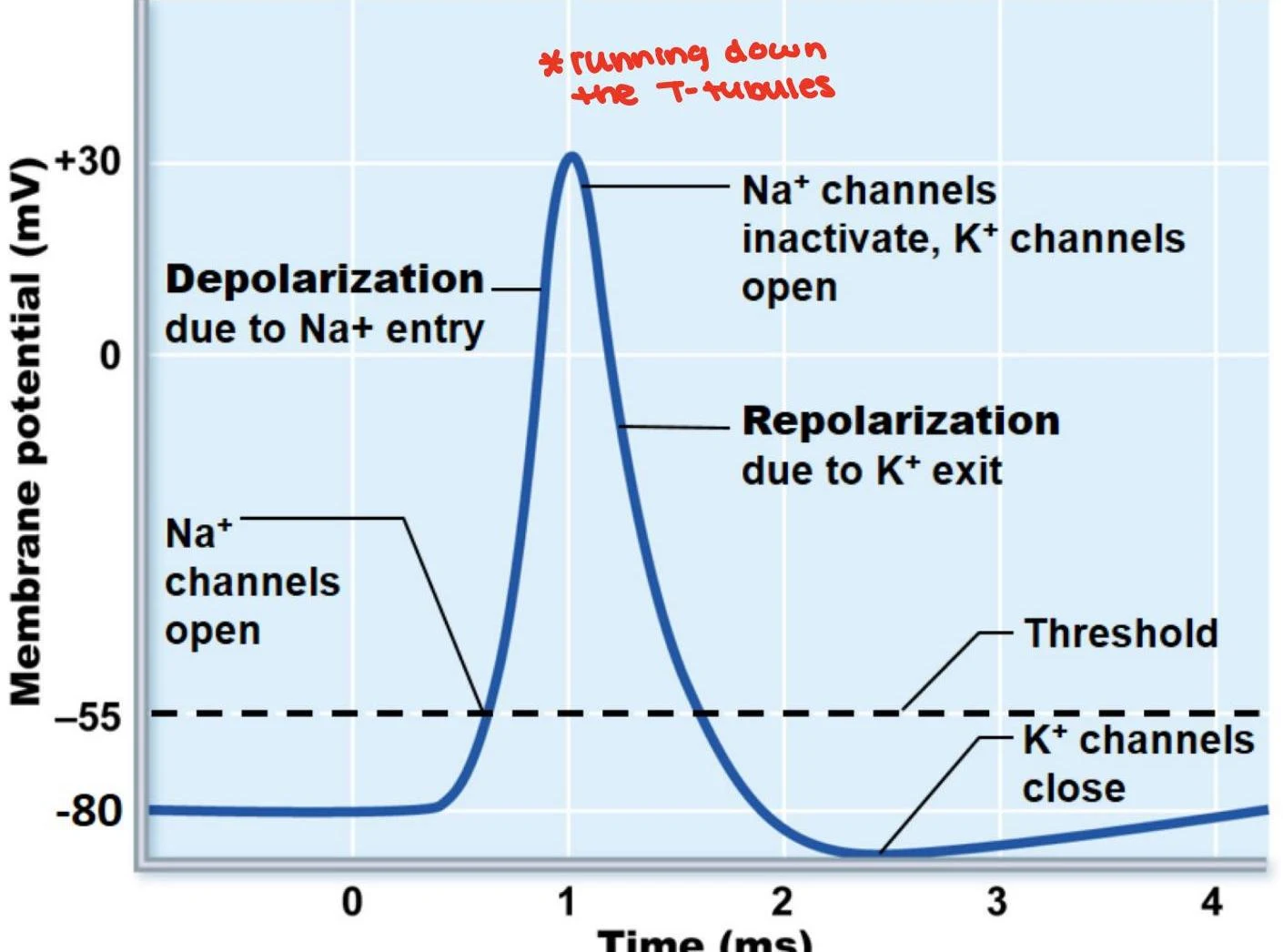
If the End Plate Potential is strong enough to reach a certain threshold, it triggers the opening of nearby voltage-gated Na+ channels, initiating a muscle action potential (AP).
The Electrical Cycle
The muscle action potential consists of three distinct phases:
Depolarization: Na+ ions rush into the cell, making the interior of the muscle cell less negative.
Repolarization: The Na+ channels inactivate, and voltage-gated $K^+$ channels open, allowing potassium to exit the cell and restoring the membrane to its initial polarized state.
Refractory Period: A brief window exists where no new action potentials can be generated until repolarization is complete, ensuring the signal moves in one direction.
This graph illustrates the changes in membrane potential during an action potential, showing depolarization due to Na+ entry and repolarization due to K+ exit.
Propagation to the Interior
The depolarization wave does not just stay on the surface; it propagates rapidly along the sarcolemma and deep into the cell via the T-tubules. This ensures that the electrical signal reaches the sarcoplasmic reticulum (SR) located deep within the muscle fiber.
Excitation-Contraction Coupling
The ultimate goal of the muscle action potential is to trigger contraction, a process known as excitation-contraction coupling.
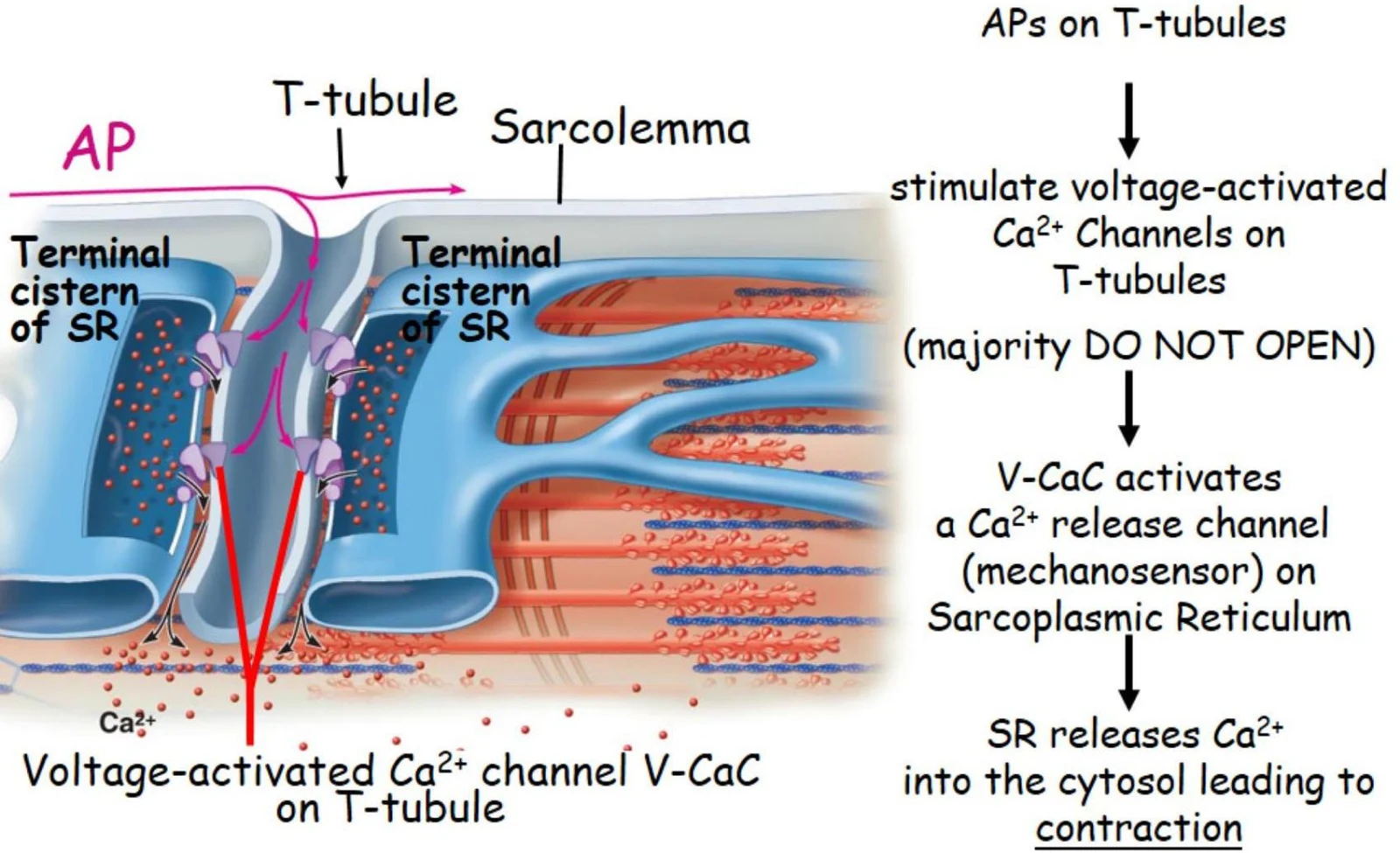
As the action potential travels down the T-tubules, it stimulates voltage-activated Ca2+ channels. These channels act as mechanosensors that trigger the release of Ca2+ from the terminal cisternae of the sarcoplasmic reticulum into the cytosol. The sudden rise in intracellular calcium is the direct signal that allows the thick and thin filaments to interact and produce force.
Diagram illustrating the process of muscle contraction, showing the role of T-tubules, sarcoplasmic reticulum, and calcium ions in initiating the process.
Exam Tip: Remember that while muscle action potentials look electrophysiologically similar to neuronal action potentials, their primary role is to act as a "trigger" for the release of calcium, rather than just transmitting information.
Speed and Frequency
Because diffusion is slower than electrical conduction, muscle cells must use high-frequency electrical impulses to maintain rapid movements. For example, physiological movements like a rattlesnake's shake or rapid muscle contractions can reach frequencies of 50 to 80 times per second.
Keep reading for free.
Create an account to finish this reading and make it part of your study routine.