


Skeletal muscle is a highly specialized tissue designed for movement, stability, and metabolic regulation. Far from being simple "rubber bands" that pull on bones, skeletal muscles are complex biological machines composed of intricate layers of connective tissue, specialized membranes, and highly organized protein filaments. Understanding how these cells function requires a deep dive into their unique cellular architecture and the electrical signals that trigger contraction.
Physiological Functions of Skeletal Muscle
Skeletal muscles are responsible for several vital physiological roles that allow for both voluntary and involuntary bodily processes.
- Movement of Bones: By pulling on bones via tendons, muscles facilitate locomotion and fine motor skills.
- Maintaining Posture and Body Position: Constant, often subconscious muscle contractions help keep the body upright and balanced. .
- Stabilizing Joints: Muscles act to bring joints back to their original state after a disturbance, providing structural stability.
- Heat Generation: Muscle metabolism consumes significant energy. When we shiver, these rapid contractions generate heat to maintain body temperature. .
Connective Tissue Organization
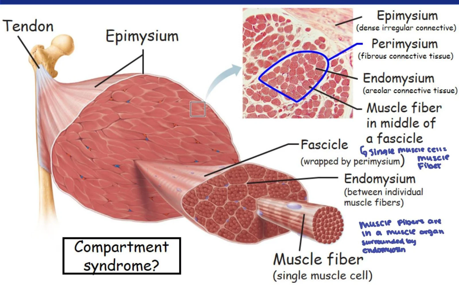
To coordinate the force generated by individual cells into a single movement, skeletal muscle is organized into layers of connective tissue. These layers ensure that the force of contraction is transmitted efficiently to the tendons and bones.
The Skeletal Muscle Fiber
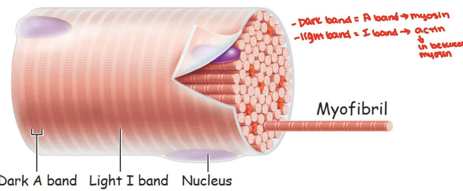
A single muscle cell is known as a skeletal muscle fiber, myofiber, or myocyte. These cells are remarkably large compared to other cells in the body, often appearing cylindrical with diameters ranging from 10 to 100 and lengths reaching up to 30 cm.



Keep reading for free.
Create an account to finish this reading and make it part of your study routine.
Continue reading freeAlready have an account? Log in
Updated