An in-depth explanation of the structure and function of skeletal muscle, covering its hierarchical organization, microscopic components, and the sliding filament theory of contraction.
Skeletal muscle is a specialized, voluntary, and striated tissue that serves as the primary engine for human movement. Beyond simple locomotion, it is essential for maintaining posture, supporting connective tissues, regulating body temperature, and storing nutrient reserves. To function effectively, all muscle cells must possess five universal properties: excitability (responsiveness to chemical or electrical signals), conductivity (the ability to propagate electrical waves), contractility (the ability to shorten), extensibility (the ability to stretch), and elasticity (the ability to recoil to a resting length).
Hierarchical Organization and Connective Tissue
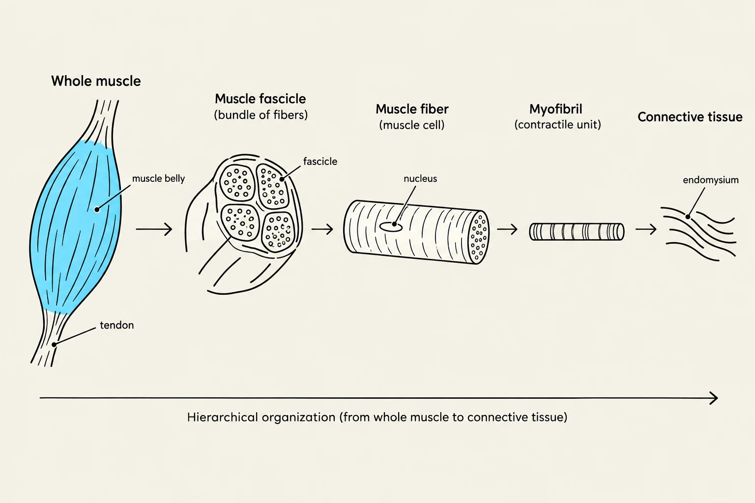
Skeletal muscle is organized in a highly structured, hierarchical manner. At the macroscopic level, a muscle is a bundle of fascicles. Each fascicle contains multiple muscle fibers, which are individual, multinucleated, cylindrical cells. Inside these fibers are long protein cords called myofibrils, which are further composed of even smaller contractile proteins known as myofilaments.
This organization is held together and protected by three distinct layers of connective tissue:
Epimysium: A layer of dense regular connective tissue that surrounds the entire muscle and separates it from surrounding organs.
Perimysium: Surrounds each fascicle and houses the blood vessels and nerves that service the muscle.
Endomysium: A thin layer of connective tissue surrounding each individual muscle fiber. This layer specifically contains satellite cells, which are embryonic stem cells vital for muscle tissue repair.
At the ends of the muscle, these three layers merge to form either a tendon or an aponeurosis to attach the muscle to bone.
This diagram illustrates the hierarchical organization of a muscle, starting from the whole muscle and breaking down to its constituent parts: muscle fascicle, muscle fiber, myofibril, and connective tissue.
The Muscle Fiber: Microscopic Components
A single muscle fiber is a complex cell characterized by its sarcolemma (the plasma membrane) and its sarcoplasm (the cytoplasm). To facilitate rapid contraction, the fiber contains specialized internal structures:
Sarcoplasmic Reticulum (SR): A specialized form of smooth endoplasmic reticulum that forms a network around each myofibril. It acts as a calcium reservoir, releasing Ca2+ to trigger contraction. Within the SR, calcium is buffered by a protein called calsequestrin, which acts like a molecular "sponge".
Transverse (T) Tubules: These are tubular infoldings of the sarcolemma that penetrate deep into the cell, allowing electrical signals (action potentials) to reach the myofibrils quickly.
Exam Tip: Remember that the T-tubules are extensions of the sarcolemma, not the SR. Their job is to carry the electrical signal from the surface into the depths of the cell so the SR can release calcium simultaneously throughout the fiber.
The Sarcomere: The Functional Unit of Contraction
The microscopic structure of a myofibril is defined by repeating segments called sarcomeres. The sarcomere is the functional contractile unit of the muscle, extending from one Z disc (or Z line) to the next. The Z disc serves as the critical anchorage point for both thin and elastic filaments.
The arrangement of myofilaments within the sarcomere creates the characteristic "striated" (striped) appearance of skeletal muscle. This pattern is composed of:
A Bands (Dark Bands): These represent the length of the thick filaments. They contain areas where thick and thin filaments overlap, as well as a central region where only thick filaments are present.
I Bands (Light Bands): These contain only thin filaments and are located spanning the Z discs.
H Zone (H Band): The central part of the A band that contains only thick filaments.
M Line: A dark protein located in the very center of the H zone.
Diagram illustrating the structure of a sarcomere, the basic contractile unit of muscle tissue, showing the arrangement of thick (myosin) and thin (actin) filaments, Z discs, and M line.
Myofilaments: The Molecular Machinery
The actual movement of a muscle is driven by the interaction of three types of myofilaments:
1. Thick Filaments (Myosin)
Myosin is a motor protein shaped like a golf club. Each molecule consists of a shaft-like tail and two globular heads. These heads form cross-bridges that reach out to bind with actin.
2. Thin Filaments (Actin, Tropomyosin, and Troponin)
Thin filaments are composed of three distinct proteins that regulate the contraction process:
F-actin: Two intertwined strands of globular (G-actin) subunits. Each G-actin has an active binding site for myosin.
Tropomyosin: A protein strand that wraps around the actin, physically blocking the active binding sites when the muscle is at rest.
Troponin: A complex of three subunits that acts as the "lock" on the system:
Troponin C: Binds to calcium ions to initiate the movement of tropomyosin.
Troponin T: Attaches the troponin complex to the tropomyosin strand.
Troponin I: Inhibits the interaction between actin and myosin to prevent unwanted contraction.
3. Elastic Filaments (Titin)
Titin is a massive, springy protein that extends from the Z disc to the M line. It acts as a "molecular spring," stabilizing the thick filaments and providing the muscle with passive elasticity, allowing it to recoil after being stretched.
The Sliding Filament Theory
Contraction does not occur because the filaments themselves shorten; rather, it occurs because they slide past one another.
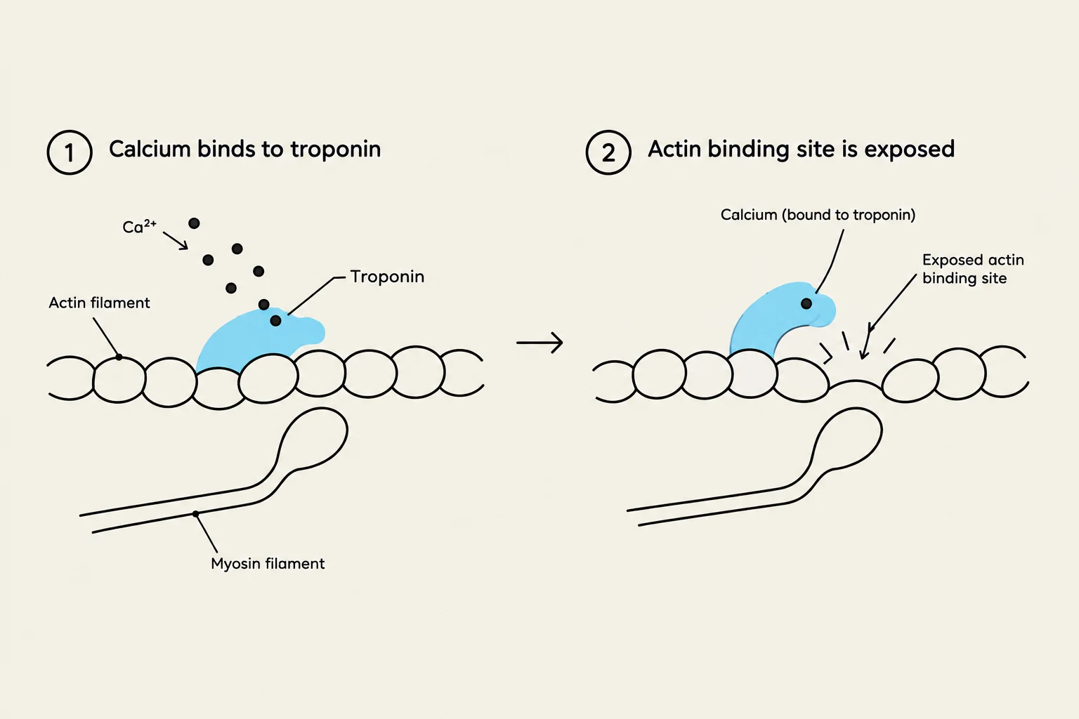
When a somatic motor neuron stimulates a muscle fiber, an action potential travels down the T-tubules, triggering the SR to release calcium into the sarcoplasm. The calcium binds to Troponin C, causing a conformational change that pulls tropomyosin away from the binding sites on the actin filament.
This initiates the Cross-bridge Cycle (or power stroke):
Cross-bridge formation: The activated myosin head binds to the exposed site on actin.
Power stroke: ADP is released, and the myosin head pivots, pulling the actin filament toward the M line.
Detachment: ATP binds to the myosin head, causing it to release its grip on the actin.
Reactivation: Myosin hydrolyzes ATP to reset its head to the "cocked" position, ready for the next cycle.
As this process repeats across thousands of sarcomeres, the Z discs are pulled closer together, the H zone and I bands decrease in width, and the overall muscle fiber shortens.
Diagram illustrating the role of calcium in muscle contraction, showing how calcium binding to troponin exposes actin binding sites for myosin interaction.
Worked Example: Calculating Sarcomere Changes
During a strong contraction, a student observes the following changes in a sarcomere:
The distance between Z discs decreases from 2.5μm to 2.0μm.
The A band width remains 1.6μm.
Question: Did the thick filaments shorten?
Answer: No. According to the Sliding Filament Theory, the A band width (which represents the length of the thick filaments) remains constant. The shortening of the sarcomere is due solely to the increased overlap of filaments.
Keep reading for free.
Create an account to finish this reading and make it part of your study routine.