


To understand how the human body moves, one must look past the skin and bone into the microscopic world of the muscle fiber. Skeletal muscle is not merely a solid mass of tissue; it is a highly organized collection of specialized proteins designed to convert chemical energy into mechanical force. By examining the internal architecture of these fibers—specifically the sarcomere, the patterns of striation, and the regulation of calcium ions—we can uncover the elegant mechanics of contraction.
The Sarcomere: The Functional Unit of Contraction
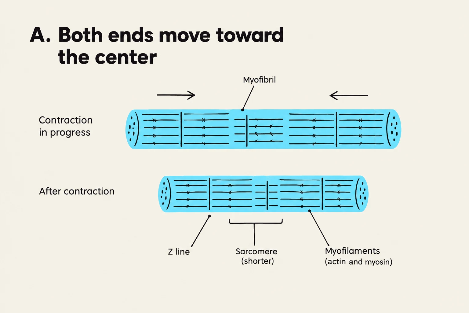
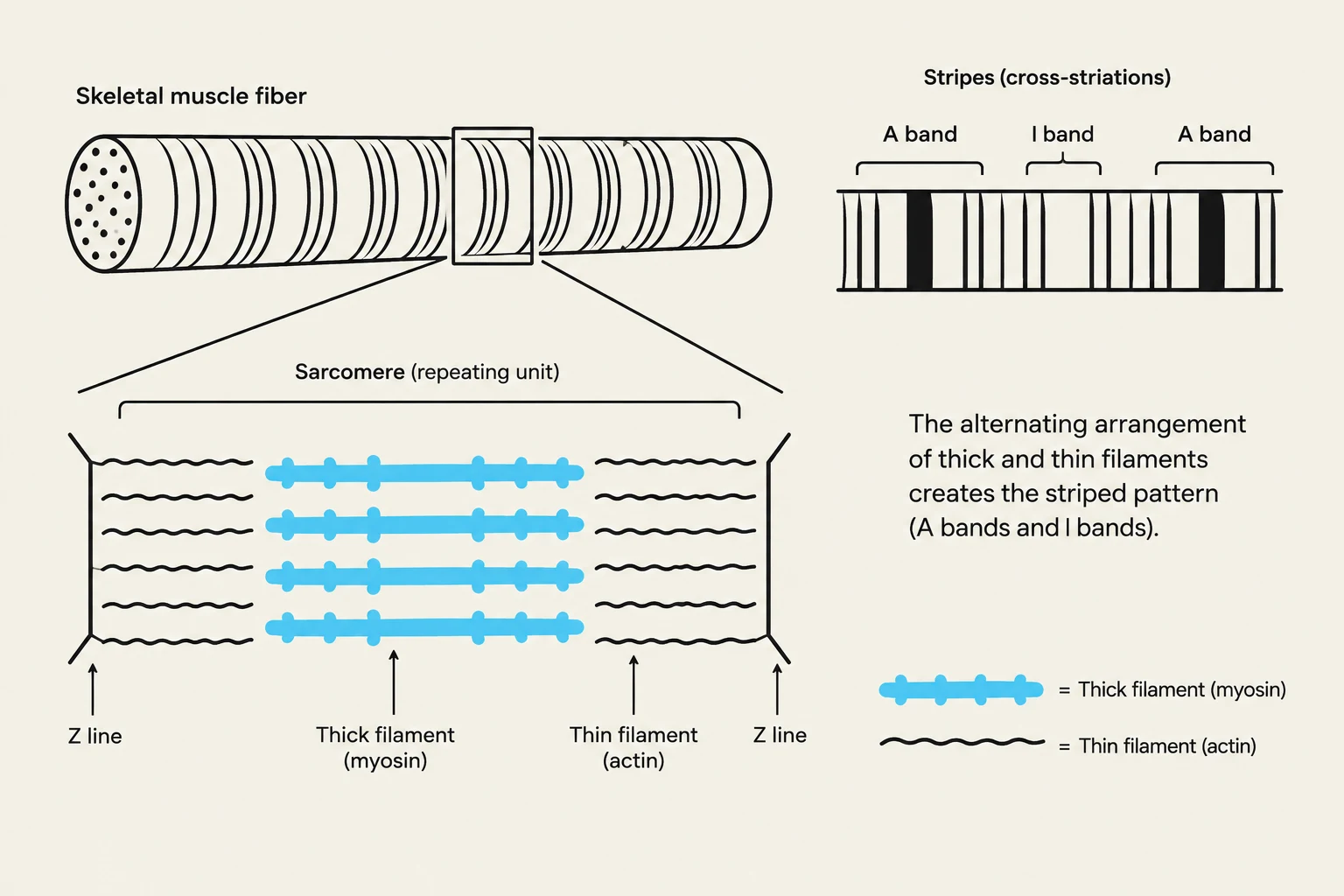
The fundamental unit of contraction in a skeletal muscle fiber is the sarcomere. A myofibril, which runs the length of the muscle fiber, is composed of many repeating segments known as sarcomeres. Within each sarcomere, the interaction between two primary types of protein filaments drives movement: the thick filaments and the thin filaments.
- Thick Filaments: Composed primarily of the protein myosin, these filaments possess "heads" that reach out to bind with the thin filaments.
- Thin Filaments: Composed primarily of the protein actin, these filaments serve as the "tracks" upon which the myosin heads pull.
The way these filaments interact depends heavily on the physical constraints of the muscle. If the ends of the myofibril are free to move, the contraction causes the ends to move toward the center. If one end is fixed (for example, attached to a bone), the free end is pulled toward that fixed point.

Keep reading for free.
Create an account to finish this reading and make it part of your study routine.
Continue reading freeAlready have an account? Log in
Updated