Propagation and Calcium Release
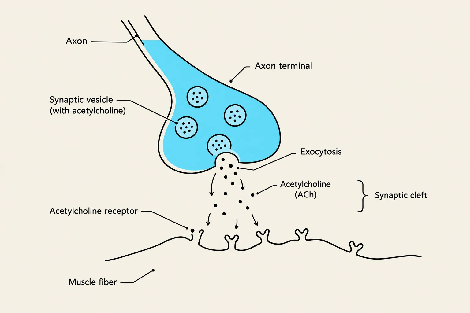
The depolarization at the NMJ triggers a new action potential that propagates along the sarcolemma and dives deep into the interior of the muscle fiber through invaginations called T-tubules (transverse tubules).
As the action potential travels down the T-tubules, it reaches the sarcoplasmic reticulum (SR), a specialized smooth endoplasmic reticulum that acts as a storage warehouse for calcium ions (Ca2+). Specifically, the signal reaches the cisterns of the SR, which are often positioned in a structure called a triad (consisting of one T-tubule and two terminal cisterns). The electrical signal causes the SR to release its stored calcium into the sarcoplasm.
Exam Tip: Remember the distinction between channel types! The calcium channels in the nerve terminal are voltage-gated, the ACh receptors on the muscle are ligand-gated, and the channels that propagate the action potential along the sarcolemma are voltage-gated.
The Contraction Cycle: Sliding Filaments
The released calcium is the "on switch" for contraction. In a resting muscle, the interaction between the thick filaments (myosin) and thin filaments (actin) is blocked by regulatory proteins: tropomyosin and troponin. Tropomyosin physically covers the binding sites on the actin filament, preventing myosin from attaching.
When Ca2+ levels rise in the sarcoplasm, the calcium binds to troponin. This binding causes a conformational change that pulls the tropomyosin away from the active sites on the actin filament, exposing them. Now, the myosin heads can bind to the actin, forming cross-bridges.
The actual movement occurs through the contraction cycle:
- ATP Hydrolysis: The myosin head binds to ATP and hydrolyzes it into ADP and inorganic phosphate (Pi), which provides the energy to "cock" the myosin head into a high-energy position.
- Cross-bridge Formation: The energized myosin head attaches to the exposed binding site on the actin.
- The Power Stroke: The release of the phosphate group triggers the "power stroke," where the myosin head pivots, pulling the actin filament toward the center of the sarcomere.
- Detachment: The release of ADP allows the head to reset, but a new molecule of ATP must bind to the myosin head to cause it to detach from the actin.
Relaxation: Resetting the System
For a muscle to relax, the stimulus must stop. First, the enzyme acetylcholinesterase (AChE) breaks down the remaining ACh in the synaptic cleft, preventing further stimulation of the sarcolemma.
Without the action potential, the calcium release channels in the SR close. To clear the sarcoplasm, active transport pumps use ATP to move Ca2+ back into the SR, where it is held by the protein calsequestrin. As calcium levels drop, the troponin-tropomyosin complex slides back into its original position, re-covering the binding sites on the actin and preventing further cross-bridge formation.
Controlling Contraction Strength
The body does not just turn muscles "on" or "off"; it modulates the intensity of the contraction through several mechanisms:
1. Motor Units and Recruitment
A motor unit is a single motor neuron and all the muscle fibers it innervates. To increase strength, the nervous system uses recruitment (multiple motor unit summation). According to the size principle, weak stimuli recruit small motor units first, while stronger stimuli recruit progressively larger units for more powerful movements.
2. Stimulus Frequency and Summation
The frequency of nerve impulses determines the duration and smoothness of a contraction:
- Muscle Twitches: A single, brief contraction and relaxation cycle.
- Temporal (Wave) Summation: If stimuli arrive in rapid succession, the twitches "ride piggyback" on one another, increasing tension.
- Tetanus: At very high frequencies, the muscle reaches incomplete (fluttering) tetanus or, if the frequency is high enough to cause a steady maximal contraction, complete (fused) tetanus.
3. Length-Tension Relationship
The amount of tension a muscle can generate depends on its initial stretch.
- If a muscle is overly stretched, there is minimal overlap between thick and thin filaments, resulting in fewer cross-bridges and weak tension.
- If a muscle is overly shortened, the filaments "butt up" against each other, also reducing force.
The nervous system maintains muscle tone—a state of partial contraction—to keep muscles near their optimum resting length for maximal force production.
Summary Table: Contraction Dynamics
| Factor | Mechanism | Result |
|---|
| Recruitment | Activating more motor units | Increased force |
| Frequency | Increasing stimulus rate | Summation → Tetanus |
| Length | Optimizing filament overlap | Maximal cross-bridge formation |