Explains the functional roles of skeletal muscles in movement, including prime movers, antagonists, synergists, and fixators. It details common movements, muscle attachments (origin and insertion), and criteria used for naming muscles. The content also covers how fascicle arrangements (circular, convergent, parallel, f
Skeletal muscles are more than just individual units of contraction; they are part of a complex, coordinated system designed to produce precise movements. Because muscles can only pull (contract) and never push, they must work in functional groups to manipulate the skeleton and move the body through space. Understanding how these muscles are organized, how they are named, and how their internal structure dictates their power is essential for studying kinesiology and anatomy.
Functional Roles in Muscle Action
When a movement occurs, muscles do not act in isolation. Instead, they participate in specific roles that allow for smooth, controlled motion.
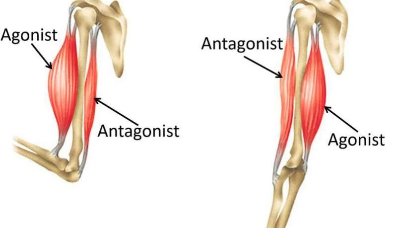
Prime Mover (Agonist): This is the muscle primarily responsible for a specific movement. For example, when you flex your elbow, the biceps brachii acts as the agonist.
Antagonist: This muscle opposes or reverses the movement of the agonist. It is typically located on the opposite side of the joint. In elbow flexion, the triceps brachii acts as the antagonist.
Synergist: These muscles assist the prime mover by adding extra force to the movement or by reducing unnecessary, unwanted movements.
Fixator: A specialized type of synergist that immobilizes a bone or the muscle's origin. By providing a stable base, the fixator allows the prime mover to act more efficiently.
Exam Tip: Remember that a single muscle can change roles! A muscle might be an agonist during one movement and an antagonist during another, depending on the direction of the limb's motion.
Diagram illustrating agonist and antagonist muscles in the human arm.
Direction of Movement and Attachments
We can often predict a muscle's function by its anatomical position relative to a joint.
Common Movements
Flexion and Extension: Muscles located on the anterior side of a joint generally cause flexion (decreasing the angle of the joint), while those on the posterior side typically cause extension (increasing the angle).
Abduction and Adduction: Muscles positioned laterally (away from the midline) typically result in abduction, while those positioned medially (toward the midline) result in adduction.
Muscle Attachments
Every skeletal muscle is defined by its attachments to other structures, such as bones or skin.
Origin: The relatively stationary, proximal attachment point.
Insertion: The distal attachment point that moves the most during contraction.
Naming Criteria for Muscles
Anatomists use several standardized criteria to name muscles, which helps in identifying their function and location:
Number of Origins: For example, biceps (two origins) or triceps (three origins).
Location of Attachments: Named by the points of origin and insertion. The sternocleidomastoid, for instance, attaches to the sternum, clavicle, and mastoid process.
Muscle Action: Named for the movement they produce, such as flexor or extensor.
Combined Criteria: Names often combine multiple descriptors. The extensor carpi radialis longus is named for its action (extensor), its attachment (carpi/wrist and radialis/radius), and its length (longus).
Fascicle Arrangements and Muscle Power
A muscle is composed of bundles of fibers called fascicles. The specific arrangement of these fascicles is a key determinant of a muscle's physical capabilities: its range of motion and its power.
Generally, there is a trade-off: long, parallel fibers allow for a greater range of motion (they can shorten more), but they are typically less powerful. Conversely, muscles with more fibers packed into a smaller area provide significant power but have a shorter range of motion.
Common Arrangement Patterns
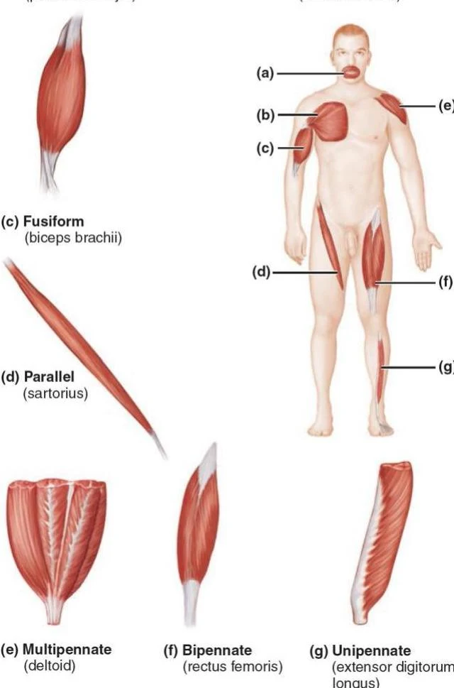
Diagrams illustrating different types of muscle fiber arrangements: fusiform, parallel, multipennate, bipennate, and unipennate, with examples of specific muscles.
Circular: Fascicles are arranged in concentric rings, often surrounding openings like the mouth (e.g., orbicularis oris).
Convergent: These muscles have a broad origin, but the fascicles converge toward a single, narrow tendon at the insertion (e.g., pectoralis major).
Parallel: Fascicles run parallel to the long axis of the muscle, creating a straplike appearance (e.g., sartorius).
Fusiform: A sub-type of parallel arrangement where the muscle is spindle-shaped, being thick in the middle and tapered at the ends (e.g., biceps brachii).
Pennate: These muscles feature short fascicles that attach obliquely (at an angle) to a central tendon. Pennate muscles are highly powerful because they can pack more fibers into a given volume. There are three types:
Unipennate: Fascicles attach to only one side of the tendon (e.g., extensor digitorum longus).
Bipennate: Fascicles insert from opposite sides of the central tendon, resembling a feather (e.g., rectus femoris).
Multipennate: Multiple directions of fascicles insert into a single tendon, appearing like many feathers (e.g., deltoid).
Summary Table: Power vs. Range of Motion
Arrangement
Fiber Length
Power
Range of Motion
Parallel/Fusiform
Long
Lower
Higher
Pennate (Bipennate/Multipennate)
Short
Higher
Lower
Clinical Note: Sarcopenia
While the study of muscle action focuses on healthy movement, it is important to note that muscle mass and function can decline with age or disease. Sarcopenia refers to the loss of muscle tissue and strength, which can significantly impact an individual's ability to perform the coordinated actions described above.
Keep reading for free.
Create an account to finish this reading and make it part of your study routine.