Explores the fundamental process of signal transduction in cells, detailing how extracellular signals are converted into intracellular responses. Covers the mechanism of signal transduction, the role of second messengers, and major types of cell surface and intracellular receptors including receptor enzymes, steroid re
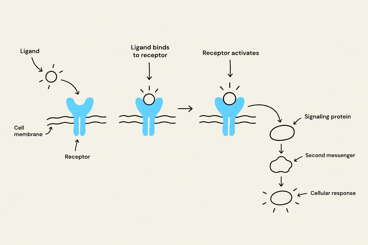
In the complex environment of a living organism, cells are constantly bombarded by chemical and physical signals from their surroundings. To survive and function, a cell must be able to sense these signals and convert them into a meaningful internal response. This process is known as signal transduction. Signal transduction involves a sequence of molecular events where an extracellular signaling molecule (the ligand) binds to a specific receptor, triggering a cascade of biochemical changes that ultimately alter the cell's behavior, such as changing its metabolism, gene expression, or movement.
The Mechanism of Signal Transduction
Signal transduction is rarely a simple one-step process. Instead, it often functions like a relay race. Once a ligand binds to a receptor, the "message" is passed through various proteins and molecules within the cell. A critical component of this relay is the use of second messengers. While the signaling molecule itself is the "first messenger," second messengers are small, non-protein, intracellular molecules (such as cyclic AMP or calcium ions) that rapidly diffuse through the cell to amplify and distribute the signal.
Exam Tip: When interpreting experimental data regarding signaling, remember that the presence of a second messenger does not always prove a direct causal link. A set of data can often lead to multiple possible conclusions, and you must design specific experiments to discern between competing hypotheses.
This diagram illustrates the process of signal transduction, where a ligand binds to a receptor on the cell membrane, initiating a cascade of events involving signaling proteins and second messengers, ultimately leading to a cellular response.
Major Types of Signal Transducers
Cells utilize different classes of receptors depending on the chemical nature of the signal they need to detect. These receptors can be broadly categorized into three major types: receptor enzymes, steroid receptors, and G-protein coupled receptors.
1. Receptor Enzymes
Receptor enzymes are transmembrane proteins where the receptor itself functions as an enzyme or is directly associated with an enzyme. When a ligand binds to the extracellular domain, it induces a conformational change that activates the intracellular enzymatic domain.
A classic example is the Receptor Tyrosine Kinase (RTK). Upon ligand binding, these receptors often dimerize (pair up), allowing the intracellular domains to phosphorylate each other on specific tyrosine residues. This phosphorylation creates docking sites for other signaling proteins, initiating a complex downstream cascade.
2. Steroid Receptors
Unlike the previous two types, steroid receptors are typically not located on the cell membrane. Because steroid hormones (such as estrogen or testosterone) are lipophilic (fat-soluble), they can diffuse directly through the lipid bilayer of the plasma membrane.
Once inside the cell, these hormones bind to steroid receptors located in the cytoplasm or the nucleus. The hormone-receptor complex then acts directly as a transcription factor, binding to specific DNA sequences to turn genes on or off. This mechanism is generally slower than membrane-bound signaling but results in long-lasting changes in the cell's proteome.
3. G-Protein Coupled Receptors (GPCRs)
G-protein coupled receptors (GPCRs) represent one of the largest and most diverse families of membrane receptors. These receptors are characterized by seven transmembrane alpha-helices that weave through the cell membrane.
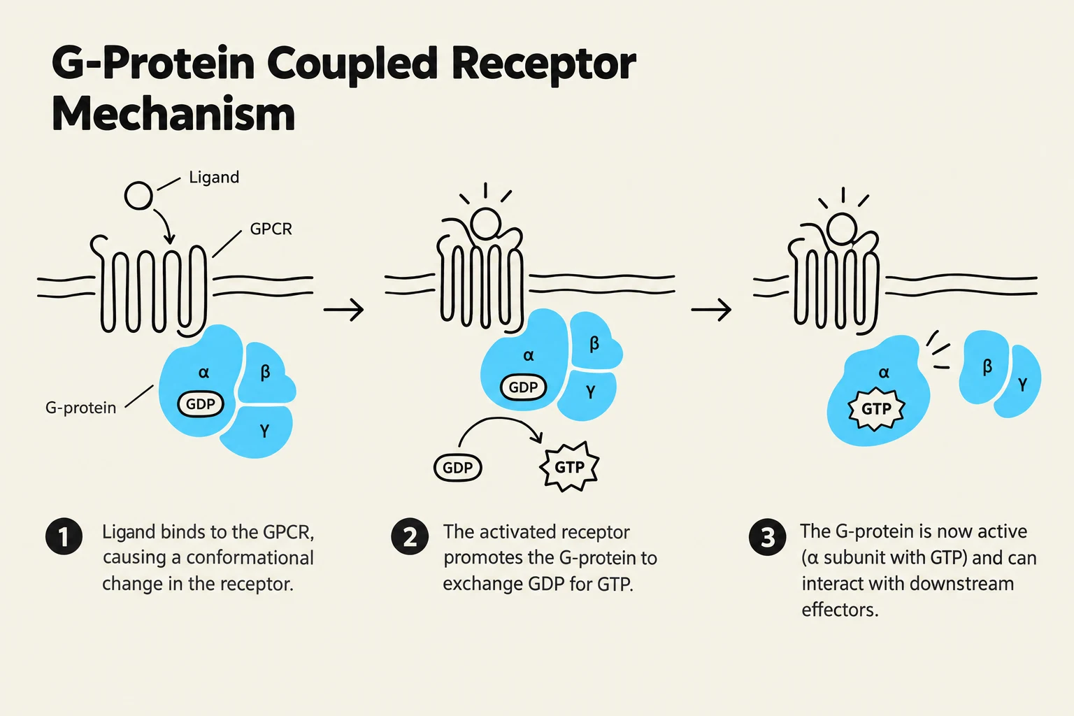
The signaling process involves G-protein linked receptors that interact with a heterotrimeric G-protein (consisting of α, β, and γ subunits). When a ligand binds to the GPCR, the receptor undergoes a conformational change that causes the associated G-protein to exchange GDP for GTP on its α-subunit. This exchange causes the G-protein to dissociate into an active α-subunit and a βγ-complex, both of which can then regulate downstream effector proteins, such as enzymes that produce second messengers.
This diagram illustrates the mechanism of G-Protein Coupled Receptors (GPCRs). It shows how a ligand binding to the GPCR causes a conformational change, leading to the activation of the G-protein by exchanging GDP for GTP. The activated G-protein can then interact with downstream effectors.
Gated Channels as Transducers
In addition to the three major types mentioned above, gated channels serve as specialized signal transducers, particularly in excitable cells like neurons and muscle cells.
Gated channels are ion channels that open or close in response to specific stimuli. These stimuli can be electrical (voltage-gated), chemical (ligand-gated), or mechanical. When a channel opens, specific ions (such as Na+, $K^+$, or Ca2+) flow down their electrochemical gradients. This rapid movement of ions changes the membrane potential of the cell, effectively transducing a chemical or electrical signal into a change in the cell's electrical state.
Summary Table of Receptor Types
Receptor Type
Location
Primary Mechanism
Example
Receptor Enzyme
Plasma Membrane
Direct enzymatic activity (e.g., phosphorylation)
Receptor Tyrosine Kinases (RTKs)
Steroid Receptor
Cytoplasm or Nucleus
Direct regulation of gene transcription
Estrogen Receptor
GPCR
Plasma Membrane
Activation of heterotrimeric G-proteins
Adrenergic Receptors
Gated Channel
Plasma Membrane
Rapid change in ion concentration/potential
Acetylcholine receptor
Keep reading for free.
Create an account to finish this reading and make it part of your study routine.