Sickle Cell Anemia and Polycythemia: An Overview of Red Blood Cell Disorders
This article provides a comprehensive overview of two significant red blood cell disorders: Sickle Cell Anemia and Polycythemia. It details the underlying mechanisms, symptoms, and classifications of each condition, highlighting the critical roles of red blood cells, hemoglobin, and blood viscosity in maintaining overa
Blood is a vital liquid connective tissue that serves as the body's primary transport system, delivering oxygen and nutrients while removing metabolic waste. Within this system, red blood cells (RBCs), or erythrocytes, play a specialized role in gas exchange using a protein called hemoglobin. However, when the composition or number of these cells becomes abnormal, it can lead to significant clinical conditions. This article explores two extremes of blood disorders: Sickle Cell Anemia, where RBCs become dysfunctional and fragile, and Polycythemia, where an excess of RBCs creates dangerous changes in blood flow.
Understanding Anemia and its Symptoms
Anemia is a condition characterized by a reduced number of RBCs or a decrease in the total hemoglobin content within the blood. Because hemoglobin is responsible for transporting oxygen, a deficiency leads to hypoxia—a state where tissues do not receive enough oxygen to support normal metabolism.
The symptoms of anemia are often a direct consequence of this oxygen deficit. Common clinical presentations include:
Fatigue and weakness: The body lacks the energy (ATP) required for physical activity due to low oxygen availability.
Dyspnea: Shortness of breath, as the body attempts to compensate for low oxygen by breathing more heavily.
Tachycardia: A rapid or irregular heartbeat as the heart works harder to circulate the limited oxygen available.
Paleness: A loss of the red color typically provided by hemoglobin.
Exam Tip: Pay close attention to specific physical signs that differentiate types of anemia. For example, koilonychia (spoon-shaped nails) is a classic clinical sign associated specifically with iron deficiency anemia.
Different types of anemia arise from different causes. Iron deficiency anemia occurs when there is insufficient iron to produce the "heme" component of hemoglobin. Pernicious anemia is an autoimmune disorder resulting from a lack of Vitamin B12, which is necessary for RBC production.
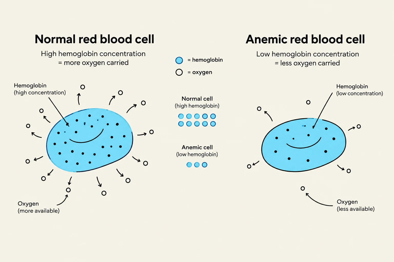
This illustration compares a normal red blood cell with high hemoglobin concentration, capable of carrying more oxygen, to an anemic red blood cell with low hemoglobin concentration, which carries less oxygen.
Sickle Cell Anemia: Mechanism and Treatment
Sickle cell anemia is a specific, severe form of anemia caused by the production of abnormal hemoglobin. In a healthy individual, hemoglobin consists of two alpha chains and two beta chains. In sickle cell patients, a mutation causes the hemoglobin to behave abnormally under low-oxygen conditions.
When oxygen levels drop, these abnormal cells lose their healthy, biconcave shape and transform into a rigid, crescent-like "sickle" shape. This leads to two major complications:
Vaso-occlusion: The rigid, sickle-shaped cells can block small arteries, leading to tissue damage, severe pain (sickle cell crisis), and potential organ failure.
Hemolysis: The abnormal cells are fragile and break down much faster than normal cells, leading to an increased destruction of RBCs. This rapid destruction can increase bilirubin levels, causing jaundice-like symptoms.
The Role of Hydroxyurea and Fetal Hemoglobin
A primary pharmacological intervention for sickle cell anemia is the drug hydroxyurea. This medication works by stimulating the body to produce more fetal hemoglobin (HbF).
Unlike adult hemoglobin, which relies on beta chains, HbF utilizes gamma chains. These gamma chains have a higher affinity for oxygen and significantly reduce the likelihood of hemoglobin polymerization (the process where hemoglobin clumps together). By increasing the proportion of HbF, the frequency of "sickling" is reduced, which in turn lowers the rates of hemolysis and vaso-occlusion.
This diagram illustrates the structural differences between adult hemoglobin (HbA), composed of two alpha and two beta chains, and fetal hemoglobin (HbF), which consists of two alpha and two gamma chains. The beta chains in adult hemoglobin are replaced by gamma chains in fetal hemoglobin.
Polycythemia: The Risks of Excess
While anemia involves too few RBCs, polycythemia is characterized by an abnormally high number of RBCs. While some individuals, such as athletes, may seek a higher RBC count to improve oxygen binding and stamina (sometimes through "blood doping"), pathological polycythemia is dangerous.
An excess of RBCs leads to two critical changes in blood composition:
Increased Hematocrit Level: The hematocrit is the percentage of total blood volume occupied by RBCs. In polycythemia, this can rise from a normal average of 45% to as high as 80%.
Increased Blood Viscosity: As the concentration of cells rises, the blood becomes thicker and more viscous. This makes blood flow slow and difficult, significantly increasing the risk of thrombosis (blood clots).
Classifications of Polycythemia
Polycythemia is categorized into two main types based on its underlying cause:
Primary Polycythemia (Polycythemia Vera): This is a bone marrow disorder where the production of RBCs is uncontrolled. It is frequently caused by a JAK-2 mutation, a genetic alteration that sends a continuous, persistent signal to the bone marrow to keep producing RBCs regardless of the body's actual needs.
Secondary Polycythemia: This is a response to external physiological factors. For example, if a person lives at high altitudes where oxygen is scarce, the kidneys sense the low oxygen (hypoxia) and release the hormone erythropoietin (EPO). EPO stimulates the bone marrow to produce more RBCs to compensate for the low oxygen levels. While this is a natural adaptation, it can also be triggered by renal tumors that produce excess EPO.
Summary Comparison Table
Feature
Anemia
Polycythemia
RBC Count
Low
High
Blood Viscosity
Low
High
Primary Risk
Hypoxia (Oxygen deficiency)
Thrombosis (Clotting/Blockage)
Key Metric
Low Hematocrit
High Hematocrit
Keep reading for free.
Create an account to finish this reading and make it part of your study routine.