Explore the biological processes of sex determination, including the roles of sex chromosomes, gamete morphology, and genetic regulation. Covers topics like phenotypic dimorphism, dosage compensation, genomic imprinting, and chromosomal abnormalities.
In the biological world, the process of establishing an organism's reproductive role is known as sex determination. While many species rely on specific genes to dictate sex, others utilize sex chromosomes—specialized, heteromorphic (dissimilar) chromosomes that distinguish the sexes. Understanding these mechanisms requires a look at how organisms differ physically, how they produce gametes, and how genetic information is regulated to ensure survival.
Reproductive Modes and Sexual Differentiation
Biological organisms exhibit a vast array of reproductive strategies. In multicellular animals, we often observe phenotypic dimorphism, where males and females look significantly different. To describe these differences, biologists distinguish between two types of development:
Primary sexual differentiation: Concerns the development of the gonads (the organs that produce gametes).
Secondary sexual differentiation: Involves the overall physical appearance and traits of the organism that are not directly part of the reproductive system.
Species are also categorized by their reproductive organs. Unisexual, dioecious, or gonochoric species possess only one type of reproductive organ (either male or female). Conversely, bisexual, monoecious, or hermaphroditic species possess both male and female organs. Some individuals may exhibit intersex characteristics, possessing intermediate traits and often being sterile.
Gamete Morphology and Sex Chromosomes
The way organisms produce gametes (sex cells) also defines their reproductive strategy. In isogamous species, such as certain fungi and algae, the male and female gametes are morphologically identical, known as isogametes. However, most eukaryotic species are heterogamous, producing morphologically distinct gametes, such as small, motile sperm and larger, nutrient-rich eggs.
In heterogamous species, the sex chromosomes determine which sex is homogametic and which is heterogametic:
Homogametic sex: Produces identical sex chromosomes (e.g., humans females are XX).
Heterogametic sex: Produces different sex chromosomes (e.g., humans males are XY).
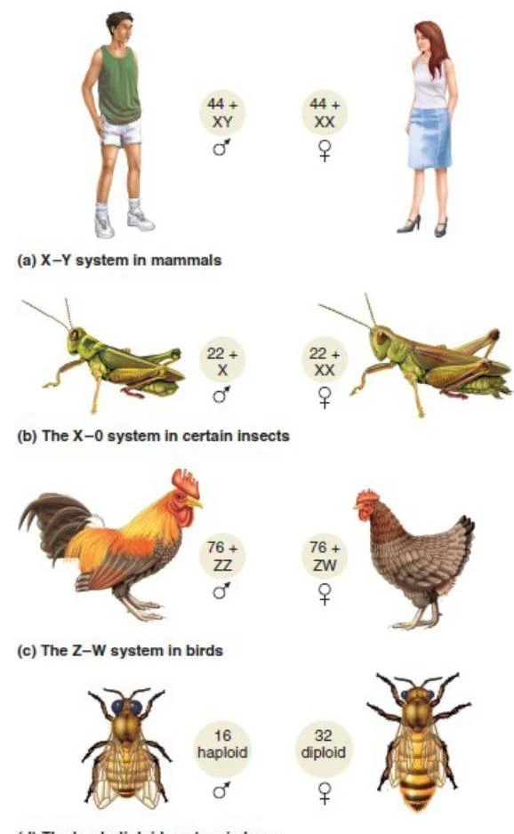
The pattern varies by species. In humans, males are the heterogametic sex (XY), but in birds (like chickens), the pattern is reversed: females are heterogametic (ZW) and males are homogametic (ZZ).
Diagrams illustrating different sex-determination systems in mammals, insects, birds, and bees.
The Human Y Chromosome and Sex Ratios
In humans, the Y chromosome is the primary determinant of maleness. It is not a uniform structure; it is divided into distinct functional regions:
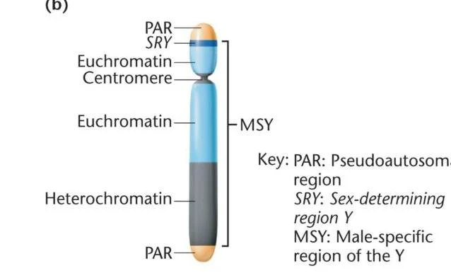
Pseudoautosomal regions (PARs): Located at the ends of the Y, these share homology with the X chromosome, allowing them to synapse and recombine during meiosis.
Male-specific region of the Y (MSY): This region does not undergo recombination with the X.
Sex-determining region Y (SRY): A critical gene within the MSY that controls male development.
Diagram illustrating the structure of the Y chromosome, highlighting the Pseudoautosomal regions (PAR), Sex-determining region Y (SRY), Euchromatin, Centromere, Heterochromatin, and the Male-specific region of the Y (MSY).
When studying populations, we distinguish between two types of sex ratios:
Primary sex ratio: The ratio of males to females at the moment of conception, which is typically approximately 1.0.
Secondary sex ratio: The ratio at birth or maturity. Interestingly, in humans, more males are born than females, likely because female embryonic mortality is higher.
Exam Tip: If you are asked to identify the genotype of a female bird, remember the ZW system. A female bird is ZW (heterogametic), while a male is ZZ (homogametic).
Dosage Compensation and the Lyon Hypothesis
Because females have two X chromosomes and males have only one, there is a potential imbalance in gene expression. To solve this, mammals use dosage compensation.
In placental mammals, this is achieved through X inactivation. According to the Lyon Hypothesis, one of the two X chromosomes in every somatic cell of a female is randomly inactivated during embryonic development. This inactivated X becomes highly condensed and is visible under a microscope as a Barr body.
Microscopic images showing a Barr body, a condensed inactive X chromosome in female cells.
The number of Barr bodies can be calculated using the formula:
Number of Barr bodies=N−1
where N is the total number of X chromosomes.
Phenotype
Sex Chromosome Composition
Number of Barr bodies
Normal female
XX
1
Normal male
XY
0
Turner syndrome
X
0
Klinefelter syndrome
XXY
1
Triple X syndrome
XXX
2
The molecular mechanism involves the X-inactivation center (Xic). The gene Xist produces a long non-coding RNA that coats the X chromosome to trigger compaction, while the gene Tsix acts as an antisense inhibitor to prevent inactivation.
Epigenetics and Genomic Imprinting
Epigenetics refers to heritable changes in gene expression that do not alter the underlying DNA sequence. A key example is genomic imprinting, where a gene's expression depends on which parent contributed the allele. This process involves chemical signatures, most notably DNA methylation, which silences genes.
Imprinting occurs in three stages:
Erasure: Existing imprints are cleared during gametogenesis.
Establishment: New imprints are set based on the sex of the individual producing the gamete.
Maintenance: The imprint is preserved in somatic cells throughout the organism's life.
Errors in imprinting can lead to severe human disorders. For example, a deletion on chromosome 15 can cause Prader-Willi Syndrome (PWS) if inherited from the father, or Angelman Syndrome (AS) if inherited from the mother.
Chromosomal Abnormalities and Variations
Genetic variation can occur through changes in chromosome structure or number.
Structural Variations
Duplication: A segment of a chromosome is repeated.
Translocation: A segment moves to a different chromosome. Reciprocal translocations can be balanced (no genetic material lost) or unbalanced (genetic material lost or gained).
Gene Families: Formed by repeated duplications, these consist of homologous genes. Related genes within a single species are called paralogs.
Copy Number Variation (CNV): Differences in the number of copies of a particular gene.
Numerical Variations
Euploidy: Possession of a complete set of chromosomes (common in plants).
Aneuploidy: An abnormal number of a specific chromosome.
Trisomy (2n+1): An extra chromosome.
Monosomy (2n−1): A missing chromosome.
Aneuploidy often results from nondisjunction, the failure of chromosomes to segregate properly during meiosis. In humans, this can lead to:
Klinefelter syndrome ($47, XXY$): Males with an extra X chromosome.
Turner syndrome ($45, X$): Females with only one X chromosome.
Mosaicism: Occurs if nondisjunction happens during mitosis, resulting in some cells having a different chromosomal makeup than others.
Extranuclear Inheritance
Finally, not all genetic information is housed in the nucleus. Extranuclear inheritance refers to DNA passed through organelles like mitochondria or chloroplasts. In many species, including humans, mitochondrial DNA is inherited exclusively from the mother.
Keep reading for free.
Create an account to finish this reading and make it part of your study routine.