Explores the biological mechanisms and pathways of gustation (taste) and olfaction (smell), detailing the cellular structures, neural transmission, and brain processing involved in these chemical senses.
Our interaction with the world is mediated by sensory pathways—complex biological routes that transmit information from the periphery to the central nervous system. While some senses, like touch, are considered general senses, others are classified as special senses because they rely on dedicated organs to detect specific stimuli. Among these special senses are gustation (taste) and olfaction (smell).
Both gustation and olfaction are categorized as chemical senses. This is because they require chemical molecules to be dissolved in a liquid medium before they can be detected by sensory receptors. For smell, volatile chemicals must dissolve in the nasal mucus; for taste, chemicals must be dissolved in saliva.
Gustation: The Sense of Taste
Taste sensation is localized within specialized structures called taste buds, which are housed in various types of protrusions on the tongue known as papillae.
The Anatomy of Papillae
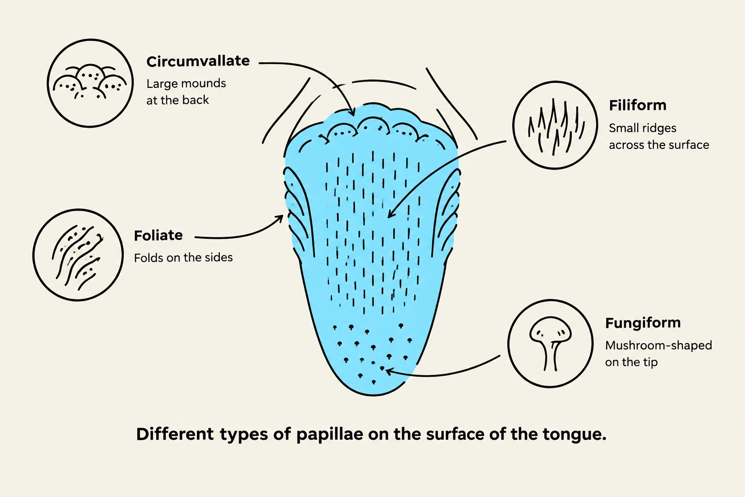
Diagram illustrating the different types of papillae on the surface of the tongue: Circumvallate, Foliate, Filiform, and Fungiform.
Not all bumps on your tongue are created equal. They serve different functions and house different amounts of sensory equipment:
Fungiform Papillae: These are small, mushroom-shaped bumps located primarily on the tip and sides of the tongue.
Foliate Papillae: These appear as folds located at the sides of the tongue.
Circumvallate Papillae: These are large, mound-shaped structures situated at the back of the tongue.
Filiform Papillae: Unlike the others, these are small ridges that provide grip and texture to the tongue but do not contain taste buds.
Cellular Composition of a Taste Bud
A single taste bud is a complex micro-environment composed of four distinct cell types:
Gustatory Receptor Cells: These contain specialized "hairs" with chemical receptor channels that detect specific molecules.
Supporting Cells: These provide structural and metabolic support for the receptor cells.
Basal Cells: Located at the base of the bud, these act as stem cells, dividing to replace both receptor and supporting cells as they age.
Gustatory Neurons: These cells associate with the receptor cells and are responsible for carrying the signal to the nervous system.
Exam Tip: Remember the distinction between papillae and taste buds. Papillae are the visible structures on the tongue, while taste buds are the microscopic sensory organs located inside specific types of papillae.
The Gustatory Pathway
This diagram illustrates the neural pathway of taste, starting from the tongue's taste receptors and progressing through cranial nerves (CN VII, IX, and X) to the brainstem, thalamus, and finally the gustatory cortex in the brain.
When chemicals trigger the gustatory receptor cells, they release neurotransmitters. These chemicals induce action potentials in the associated gustatory neurons. The transmission of this information to the brain is handled by three specific cranial nerves:
CN VII (Facial Nerve): Carries taste from the anterior (front) two-thirds of the tongue.
CN IX (Glossopharyngeal Nerve): Carries taste from the posterior (back) third of the tongue.
CN X (Vagus Nerve): Carries taste sensation from the back of the throat.
Once the signal leaves the cranial nerves, it follows a specific trajectory through the brain:
Brainstem: The signal first reaches the brainstem (specifically the medulla oblongata and pons).
Thalamus: The signal is sent to the thalamus, which acts as the central relay and integration hub for sensory information.
Cerebral Cortex: Finally, the information reaches the cerebral cortex (specifically the gustatory cortex) for conscious perception and processing.
Olfaction: The Sense of Smell
Olfaction follows a cellular logic similar to gustation but utilizes a different anatomical structure: the olfactory epithelium.
Cellular Structure of the Olfactory Epithelium
The olfactory epithelium contains specialized cells that facilitate the detection of volatile chemicals:
Olfactory Receptor Cells: These possess dendrite-like branches that extend toward the surface to capture chemical stimuli.
Supporting Cells: These cells provide physical support and are responsible for secreting the mucus necessary to dissolve odors.
Basal Cells: Similar to taste buds, these serve as stem cells to replace damaged receptors and supporting cells.
Olfactory Neurons: These cells receive neurotransmitters from the receptor cells to initiate the neural signal.
The Mechanism of Detection
When an odorant molecule reaches the epithelium, it triggers a chemical change in the receptor cell. Specifically, the stimulus causes Na+ (sodium) to enter the cell, leading to depolarization. This change in voltage triggers the release of neurotransmitters, which then induce action potentials in the olfactory nerve cells.
Olfactory Pathways and Emotional Connection
The olfactory pathway is unique in its ability to link directly to areas of the brain responsible for memory and emotion:
The Olfactory Bulb: The initial processing center for smell.
The Amygdala: A deep brain region that processes emotions, fear, and survival responses.
The Hippocampus: A structure in the temporal lobe essential for learning, memory formation, and spatial navigation.
This connection explains why certain smells can trigger intense, sudden memories or emotional reactions.
Summary Comparison of Chemical Senses
Feature
Gustation (Taste)
Olfaction (Smell)
Medium Required
Saliva
Nasal Mucus
Primary Organ
Tongue (Papillae/Taste Buds)
Olfactory Epithelium
Key Nerve(s)
CN VII, CN IX, CN X
CN I (Olfactory Nerve)
Relay Center
Thalamus
Thalamus / Olfactory Bulb
Note on Equilibrium: While taste and smell are chemical senses, the body also maintains equilibrium (balance and spatial orientation) through the vestibular system in the inner ear. This is a separate special sense that does not rely on chemical detection.
Keep reading for free.
Create an account to finish this reading and make it part of your study routine.