Sensory and Motor Pathways and Reflexes in the Human Body
Explores the communication loop between the peripheral and central nervous systems, detailing sensory input pathways, motor output pathways, and the role of reflexes in rapid responses.
The human body relies on a continuous, high-speed communication loop between the peripheral nervous system (PNS) and the central nervous system (CNS). This loop allows us to perceive our environment through sensory input and respond to it through coordinated movement. This article explores the complex pathways that carry information to the brain and the motor commands that return to our muscles, as well as the rapid, automatic responses known as reflexes.
Sensory Pathways: From Receptors to the Cortex
The sensory pathway from receptor to somatosensory cortex.
Sensory information begins at specialized receptors in the periphery. These receptors convert physical or chemical stimuli into electrical signals that travel along afferent pathways.
Mechanoreceptors and Proprioceptors
The general senses rely on various types of receptors to interpret the world. Mechanoreceptors are essential for tactile sensations, detecting stimuli such as light touch, pressure, temperature, and vibration.
Beyond simple touch, the body utilizes a specialized group of receptors called proprioceptors to maintain awareness of body position and movement. Proprioception is vital for activities like walking or reaching for an object without looking. These receptors include:
Muscle Spindles: These detect the stretch of skeletal muscles.
Tendon Organs: These monitor the stretch of tendons during muscle contractions.
Synovial Joint Receptors: These relay information regarding the movement and position within the joints.
Processing the Input
Once a stimulus is detected, the afferent neurons carry the signal toward the spinal cord and up to the brain. Most sensory information is routed through the thalamus, which acts as a relay station, before being sent to the somatosensory cortex located in the parietal lobe of the brain.
In some cases, sensory information is transmitted via cranial nerves directly to specific brain regions. For example, olfactory (smell) information travels via the olfactory nerve to the olfactory bulb and cortex.
Exam Tip: Remember the distinction between "Afferent" and "Efferent." Afferent pathways Arrive at the CNS (Sensory), while Efferent pathways Exit the CNS (Motor).
Motor Pathways: Coordinating Movement
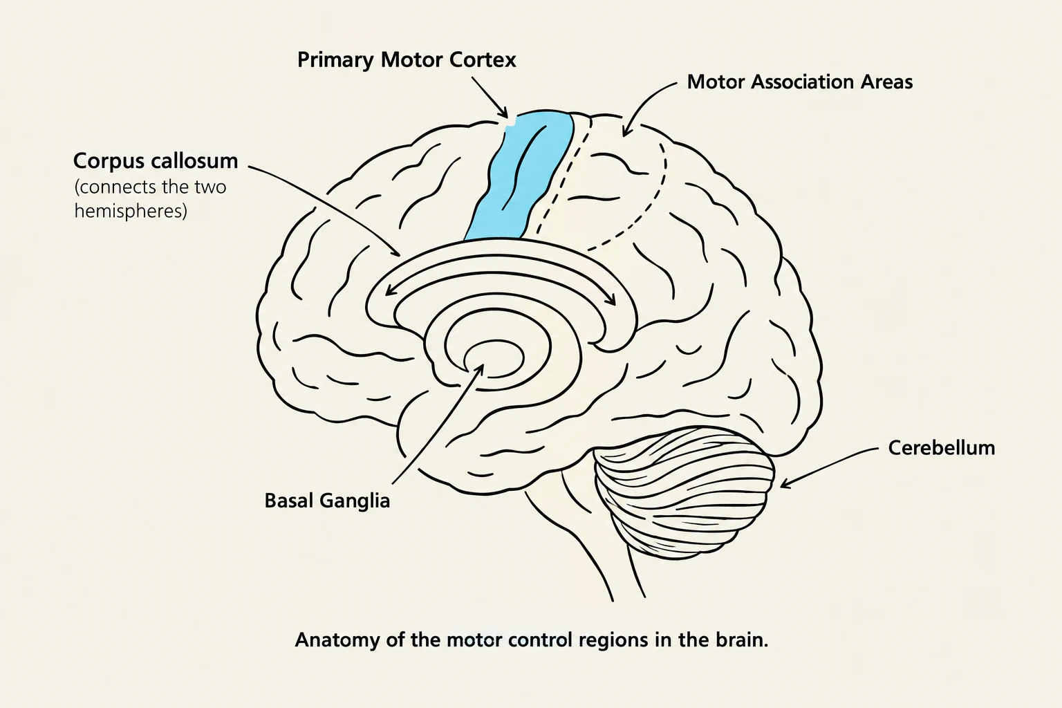
An illustration showing the anatomy of the motor control regions in the brain, including the primary motor cortex, motor association areas, corpus callosum, basal ganglia, and cerebellum.
Once the brain processes sensory data, it must send instructions back to the body to initiate action. This is the role of the efferent pathways, which carry motor information from the CNS to effector organs, such as muscle cells.
Key Brain Regions for Motor Control
Motor control is not managed by a single "button" in the brain; rather, it is a highly coordinated effort involving several specialized structures:
Primary Motor Cortex: The specific cortical region responsible for executing voluntary movements.
Motor Association Areas: These regions are responsible for the higher-level "planning" phase, including the sequencing of complex movements.
Cerebellum: This structure acts as the body's quality control, coordinating balance, precision, and timing of movement.
Basal Ganglia: These structures help regulate voluntary movements by filtering signals, ensuring that intended movements occur smoothly while unwanted movements are suppressed.
Cerebral Nuclei: These clusters of neuronal cell bodies are involved in broader motor control, learning, and executive functions.
To facilitate communication between the two hemispheres of the brain, the corpus callosum—a thick band of nerve fibers—allows for the integration of motor information across the left and right sides of the body.
Peripheral Communication: The Role of Plexuses
For motor commands to reach the limbs, they must pass through complex networks of nerves known as plexuses. These networks facilitate the communication between the spinal cord (CNS) and the limbs (PNS).
The three major plexuses include:
Brachial Plexus: Connects the spinal cord in the neck to the shoulders, arms, and hands.
Lumbar Plexus: Connects the spinal cord in the lower back to the abdominal wall, external genitalia, and parts of the lower limbs.
Sacral Plexus: Connects the spinal cord in the lower back to the buttocks, legs, feet, and parts of the pelvis.
Worked Example: The "Crumb" Scenario
Imagine you have a crumb on your lip.
Sensory Phase: Mechanoreceptors on your lip detect the particle. Sensory nerves (such as the Trigeminal or Facial cranial nerves) transmit this via afferent pathways to the brain.
Integration Phase: The brain processes the location and decides to move the tongue.
Motor Phase: The brain sends signals through efferent pathways. The Hypoglossal nerve (CN XII) triggers tongue movement, while the Trigeminal nerve (CN V) might trigger jaw movement to help clear the crumb. If you need more complex movement, like lifting your shoulder, signals may travel through the brachial plexus.
Reflexes: Rapid Response Systems
Comparison of monosynaptic and polysynaptic reflex arcs, illustrating the neural pathways involved in simple and complex reflexes.
While most sensory information requires conscious processing in the cortex, some stimuli require an immediate, automatic response to protect the body. These are known as reflexes. Reflexes are categorized by how many synapses (connections between neurons) occur within the spinal cord:
Monosynaptic Reflexes
A monosynaptic reflex is the simplest form of a reflex. It involves only a single synapse within the spinal cord, allowing for direct, lightning-fast communication between a sensory neuron and a motor neuron.
Example: The stretch reflex (such as the knee-jerk reflex) is a classic monosynaptic response.
Polysynaptic Reflexes
A polysynaptic reflex involves two or more synapses within the spinal cord. Because the signal must pass through one or more interneurons before reaching the motor neuron, the communication is "indirect" and slightly slower than a monosynaptic reflex.
Example: The withdrawal reflex (pulling your hand away from a hot stove) is a polysynaptic reflex.
Keep reading for free.
Create an account to finish this reading and make it part of your study routine.