


Imagine you are embarking on a long journey through the wilderness on a bicycle. To ensure you don't get stranded, you pack a repair kit: spare tires, a patch kit, and tools to fix a flat. In the human body, skeletal muscle possesses a remarkably similar built-in system. Every individual muscle fiber comes equipped with its own personal "repair kit" located right next to it, tucked underneath the basal lamina. This specialized repair kit is composed of satellite cells.
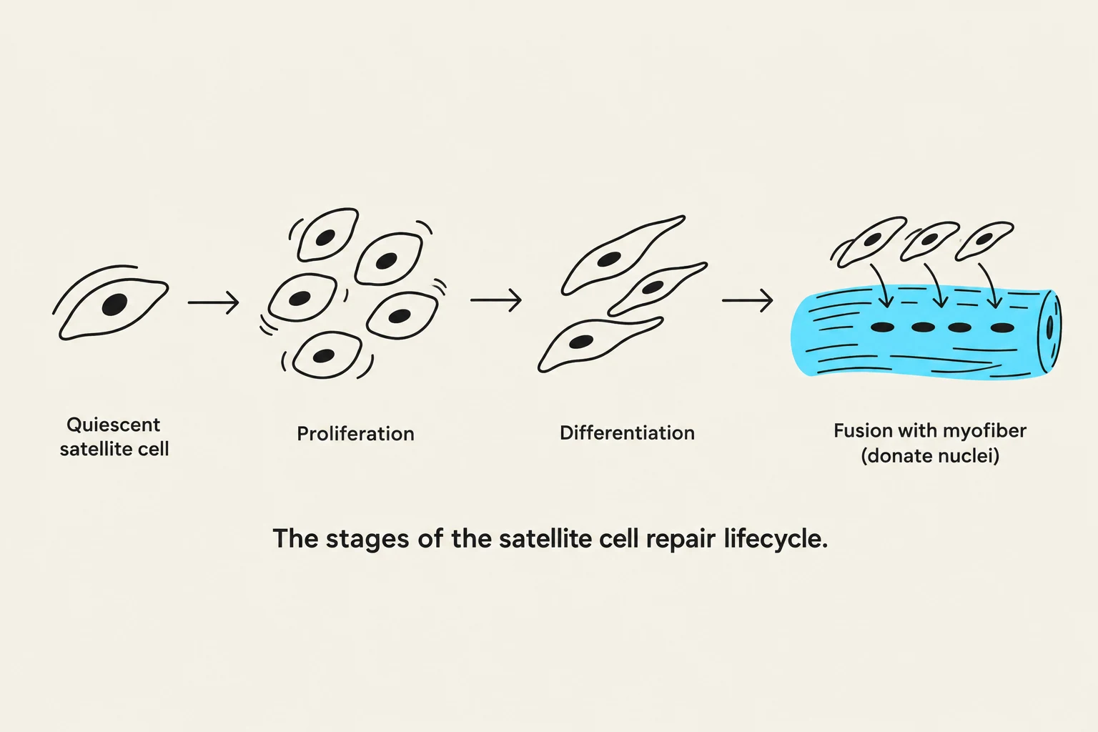
Because mature muscle fibers (myofibers) are unable to divide to create new cells, they cannot repair themselves through traditional cell division. Instead, they rely on the activation of these specialized progenitor cells to facilitate both the repair of damaged tissue and the growth of muscle mass.
The Anatomy of a Myofiber
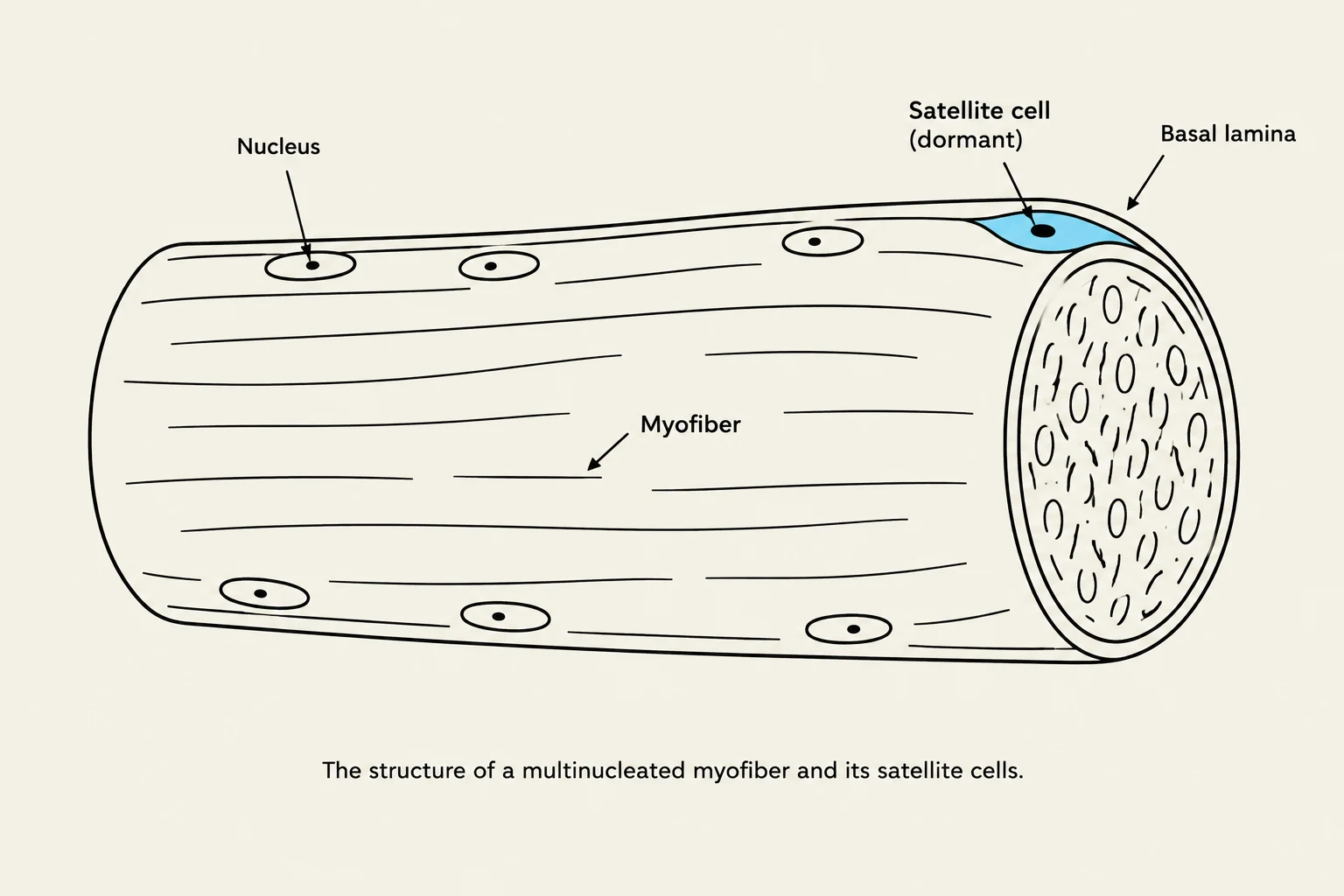
To understand how repair works, we must first understand the structure of the cell being repaired. A muscle is composed of many individual myofibers. Unlike most cells in your body, a myofiber is multinucleated, meaning it contains many nuclei distributed along its length.
These nuclei are essential because they act as the control centers for the cell. In a large, complex structure like a myofiber, a single nucleus cannot manage the entire cell; instead, each nucleus controls a specific "domain" or region of the fiber, overseeing the synthesis of proteins necessary for maintaining and expanding the muscle.
Exam Tip: Identifying Nuclei In histological images (like those using DNA stains), muscle nuclei are typically located on the periphery of the cell, pushed against the membrane to make room for the dense contractile machinery inside.

Keep reading for free.
Create an account to finish this reading and make it part of your study routine.
Continue reading freeAlready have an account? Log in
Updated